| ORIGINAL ARTICLE |
|
|
1 The First Affiliated Hospital of Shantou University Medical College, Shantou 515041, China;
2 Brain Function and Disease Laboratory, Shantou University Medical College, Shantou, 515041, China
Corresponding Author: Xiaoyu Ji, The First Affiliated Hospital of Shantou University Medical College, Shantou 515041, China; Brain Function and Disease Laboratory, Shantou University Medical College, Shantou, 515041, China. E-mail: xyji@stu.edu.cn.
Running title: STUDY ON EFNA3 IN HEPATOCELLULAR CARCINOMA
| |
ABSTRACT |
| INTRODUCTION | |
|
|
MATERIALS AND METHODS |
|
|
RESULTS |
|
|
DISCUSSION |
|
|
CONCLUSION |
|
|
AUTHOR CONTRIBUTIONS |
|
|
FUNDING |
|
|
COMPETING INTEREST |
|
|
ETHICS APPROVAL AND CONSENT TO PARTICIPATE AND PUBLISH |
|
|
REFERENCES |
|
|
ABSTRACT
|
|---|
Hepatocellular carcinoma (HCC) is a common disease. Multitarget therapy has been considered to be effective. EphrinA3 (EFNA3) ligand has been proven to be related to many cancers but is rarely reported in HCC. The purpose of this study was to investigate the clinical significance and mechanism of EFNA3 in HCC. EFNA3 ligand has been related to many cancers. By analyzing data from the gene expression omnibus (GEO) and The Cancer Genome Atlas (TCGA) databases with R software, EFNA3 was found highly expressed in patients with HCC, and the group with higher EFNA3 expression in patients with HCC had a significantly poorer prognosis. EFNA3 expression was of high diagnostic value and can be an independent clinical prognostic factor in HCC according to the COX regression and the area under the receiver operating characteristic (ROC) curve. Anomalies in the EFNA3 gene suggested a poor prognosis in patients with HCC analyzed by cBioPortal. The expression of EFNA3 was positively correlated with the infiltration of immune cells and two immune checkpoints. The function of EFNA3 and its related genes has been linked to the Ras signal pathway and affected the development of HCC. Based on exploring the clinical value of EFNA3, this study used the bioinformatics method to reveal the role of EFNA3 in immune regulation and its possible mechanism in hepatocellular carcinoma.
KEY WORDS: EFNA3; hepatocellular carcinoma; biomarker; gene expression; immune cells|
|
INTRODUCTION |
|---|
Liver carcinoma is a frequent cancer form, ranking sixth among the most common type of tumors globally and the third major cause of tumor fatality. There were around 906,000 new cases and 830,000 deaths globally in the year 2020 and HCC or liver hepatocellular carcinoma (LIHC) is the highest incidence type among them, contributing to approximately 75% to 85% of cases (1). Because of the limited effect of treatment methods, for example, surgery is the main treatment scheme and is almost only suitable for early HCC. Still, the probability of metastasis and recurrence after surgery is high. HCC has a dismal prognosis (2, 3).
Nowadays, immunotherapy has become a compelling way to tackle the disease. By forming a symbiotic interaction with cancer cells, the immune microenvironment of HCC influenced tumor evolvement, invasion, recrudescence, and metastasis (4, 5), the most effective treatments may entail attacking various identified targets (6), Thus it is necessary to continue exploring therapeutic targets of HCC to find a more effective treatment, provide patients with scientific and reasonable treatment, and improve their prognosis.
The signal transduction pathway of receptor tyrosine kinase (RTK) is critical in developing HCC (7). It was reported that RTK plays an essential role in the cell growth process, motility, survival, and differentiation (8). Erythropoietin-producing hepatocellular (Eph) receptor interacting with plasma-membrane-bound ephrin ligands is one of the major subsets of the RTK family iso be relevant to tumors (9). There are currently 14 Eph receptors and 8 Ephrin ligands discovered in humans. EF-NA3 is one of the Ephrin ligands and an explicit miR-210 target (10). It can inhibit angiogenesis. MiR-210 can regulate cancer cell propagation, metastasis, and apoptosis (11). MiR-210 induces migration and angiogenesis in endothelial cells by inhibiting EFNA3 (10). At the same time, some studies have shown that it is possible to mediate the high expression of EFNA3 in cancer through a long-chain non-coding RNA, which was related to the hypoxia setting (12).
In recent years, it has been proved that the Eph/Ephrin signaling pathway composed of EFNA3 and its receptor is related to many tumors' occurrence, development, and prognosis. Such as gastric cancer [13] and lung adenocarcinoma (14). Given the role of EFNA3 in these tumors, it is necessary to further explore EFNA3 in HCC patients. However, there are few studies about the expression of EFNA3 in HCC. In this research, we applied the TCGA database (https://portal.gdc.cancer.gov, a milestone dis-ease genomics program, microscopically portrayed north of 20,000 primary malignant growth and matched typical examples traversing 33 malignant growth types) to investigate the diagnostic and prognostic value of EFNA3 expression in HCC and validated with two GEO datasets, as well as exploring its association with immune infiltration, biomarkers, and immunological checkpoints by various tools. The mechanism involved in the incidence and progression of HCC, containing gene alternation and enriched signalling pathways was investigated.
|
|
MATERIALS AND METHODS |
|---|
Expression analysis of EFNA3 in primary tumors and paired normal tissue
We use UCSC Xena (https://xenabrowser.net/datapages) to uniformly handle TCGA and the Genotype-Tissue Expression (GTEx, https://www.gtexportal.org, continuous work to construct an extensive public asset to concentrate on tissue-specific gene expression and regulation) RNAseq TPM format data and compare analysis after log2 transformed.
EFNA3 differential expression in HCC patients
Through the TCGA database, we downloaded TCGA HCC Patient RNA-Seq expression profile data (424 samples including 50 normal and 374 tumor tissues, Workflow Type: HTSeq-FPKM) and clinically relevant data in March 2022. We compared these RNA-Seq with log2 logarithms, and we compared 50 cancer samples with matched normal samples to make the results more reliable. Meanwhile, immunohistochemical data of HCC patients and normal liver tissues were gained for verification from the Human Protein Atlas (HPA, https://www.proteinatlas.org, being committed to providing all Human proteins of tissue and cell distribution information).
HCC clinical correlation analysis and survival analysis of EFNA3
The relationship between EFNA3 and clinical characteristics was analyzed by R package ggplot2. The Kaplan-Meier (KM) plot was utilized to show the prognosis of HCC patients on the overall survival (OS) with various degrees of EFNA3 expression by the survminer package and survival package with R. Cox regression methods were performed to investigate the effect of EFNA3 on the overall survival (OS) of HCC by the R package survival. We estimated the diagnostic value of EFNA3 by utilizing the area under the ROC curve with R packages ggplot2 and pROC. Finally, by combining EFNA3 with clinical variables, patients' 1-, 3-, and 5-year survival probabilities were predicted using a nomogram with the R packages survival and rms. The above analysis was all based on the TCGA database.
Validation with GEO database
From the GEO database (https://www.ncbi.nlm.nih.gov/geo, including research institutions around the world to submit high-throughput gene expression data), we further verified the reproducibility of EFNA3 expression differences by comparing log2 logarithms of RNA-seq of these samples and detected diagnostic value in HCC patients with the area under the curve of ROC in GSE14520 and GSE76427 datasets.
Gene alteration in patients with HCC
On cBioPortal (https://www.cbioportal.org), the alternation of the EFNA3 gene was analyzed for three data sets of HCC (TCGA, Firehose Legacy; AMC Hepatology 2014; INSERM, Nat genet 2015). To examine the significance of the difference in survival, KM plots and a log-rank test were performed.
Correlation analysis of EFNA3 with immune cell infiltration and immune checkpoints
TIMER (http://timer.cistrome.org) online tool was utilized to investigate the relationship between EFNA3 expression and immune infiltration in HCC. In addition, the correlation of EFNA3 with the immune checkpoint was investigated.
Enrichment analysis using Gene Ontology (GO) and the Kyoto Encyclopedia of Genes and Genomes (KEGG)
The STRING database (https://string-db.org) was used to gain protein-protein interaction (PPI) network information of EFNA3. Protein interaction was considered statistically significant when the interaction score was greater than 0.9. A total of seven EFNA3 functional partner genes were used in GO and KEGG enrichment analyses with R packages clusterProfiler and org.Hs.eg.db.
Data analysis
R (3.6.3) software was utilized to analyze the data downloaded from the TCGA database. To compare the differences between groups, the student’s t-test or Wilcoxon rank-sum test was conducted. Correlations were examined using Spearman or Pearson correlation tests appropriately. The difference in prognosis was determined by log-rank tests. The importance of single factor cox analysis (P<0.1) was incorporated in the multivariate analysis in the Cox regression analysis. Statistical significance was defined as a P-value of less than 0.05.
|
|
RESULTS |
|---|
Expression analysis of EFNA3 in primary tumors and paired normal tissue
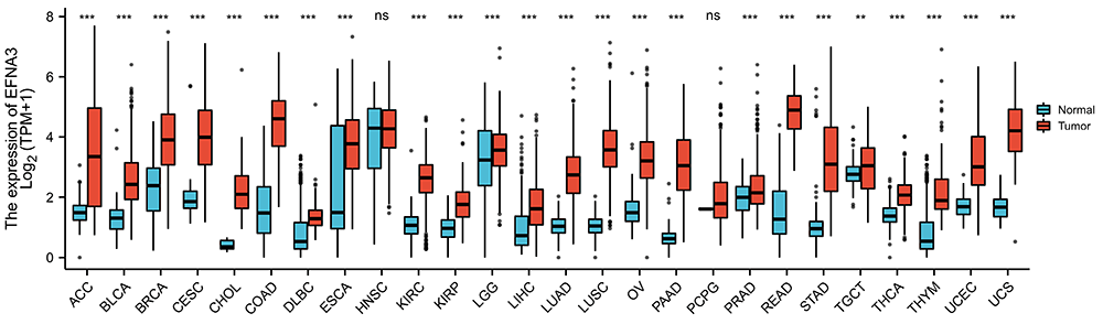
To investigate the potential roles of EFNA3, the expression of EFNA3 in 26 types of cancer in human was compared with homologous normal tissue (Figure 1). In breast invasive carcinoma (BRCA), kidney renal clear cell carcinoma (KIRC), liver hepatocellular carcinoma (LIHC), lung adenocarcinoma (LUAD) and other cancers, the expression level of EFNA3 in cancer tissues was significantly higher than that in normal tissues. However, no difference was found in head and neck squamous cell carcinoma (HNSC) and pheochromocytoma and paraganglioma (PCPG).
Higher EFNA3 expression in HCC patients than normal liver tissue
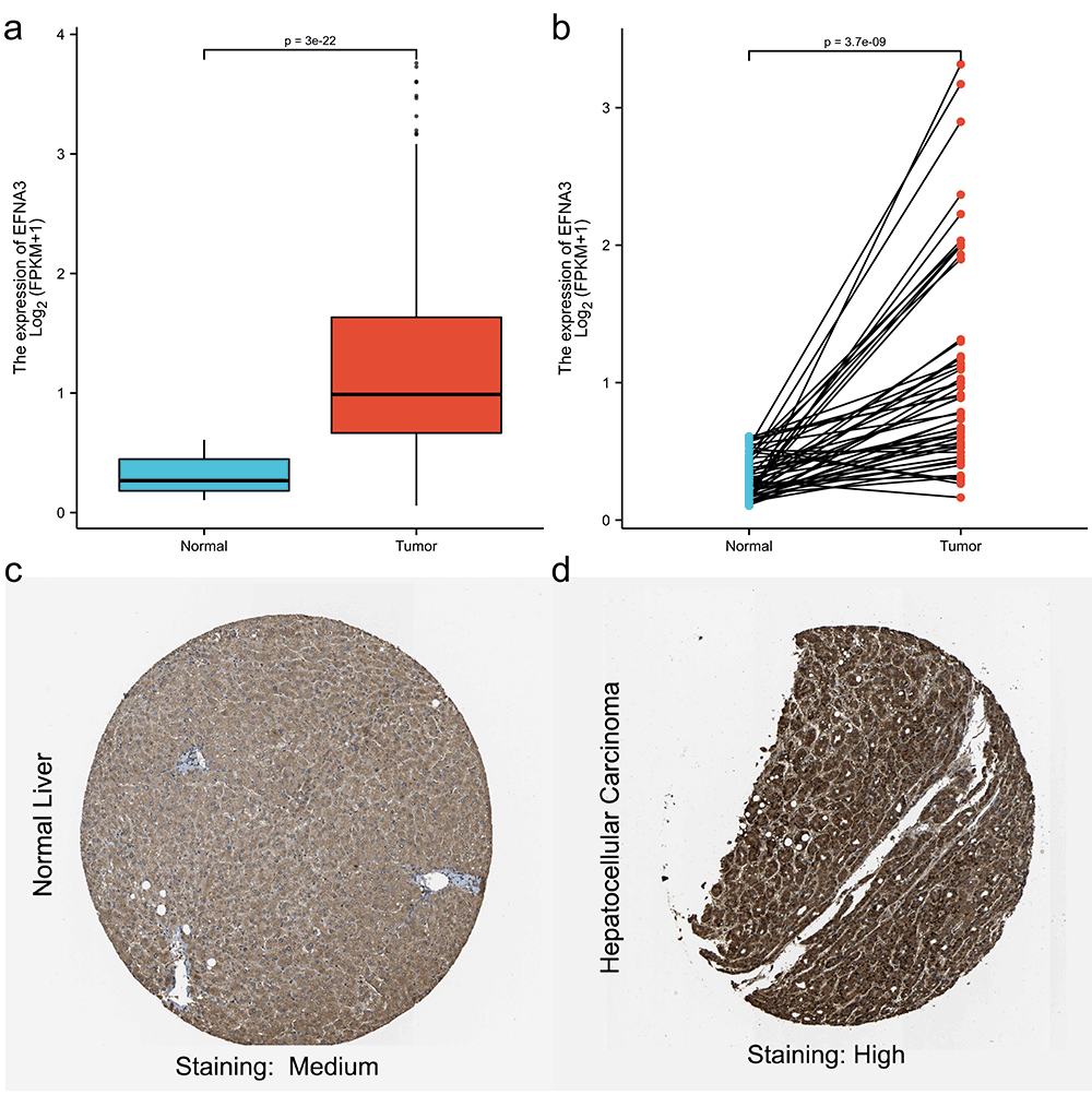
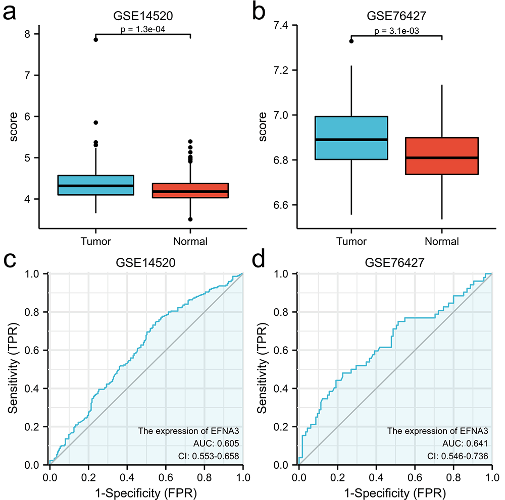
The expression levels of EFNA3 in HCC patients were compared to those in normal liver tissues to determine its status. The expression of the EFNA3 gene was discovered to be substantially higher in HCC tissues compared with that in normal tissues (P<0.001 in Figure 2a). This finding was confirmed in paired HCC tissues paired with normal liver tissues (P<0.001 in Figure 2b). Immunohistochemical staining samples of HCC and normal tissues were obtained from the HPA database, and it was discovered that EFNA3 expression in HCC was substantially higher than in normal tissues (Figure 2c, d). In the GSE14520 dataset, we found that EFNA3 was also highly expressed in hepatocellular patients (P<0.05, Figure 5a, b).
Clinical correlation analysis of EFNA3 in HCC
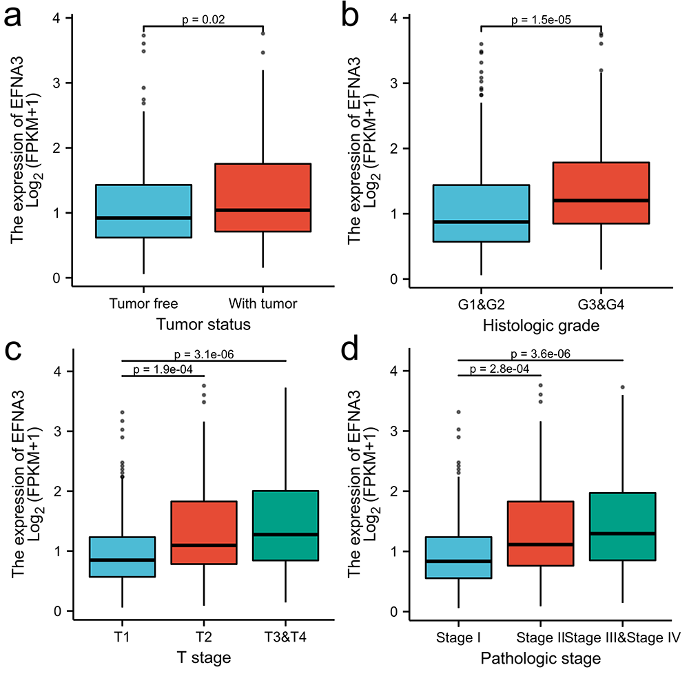
Tumor status (P=0.02), histologic grade (P=1.1e-05), pathologic stage (P=4.6e-06), and T stage (P=3.9e-06) had significant differences in EFNA3 expression (Figure 3a-d). Logistic analysis of EFNA3 expression level and clinical characteristics showed that high EFNA3 expression was intimately correlated with race, tissue grade, pathological stage, and T stage (P<0.001 in Table 1), whereas EFNA3 expression did not differ significantly in clinical characteristics such as age, gender, N stage, and M stage.
Prognostic analysis and diagnostic value of EFNA3 in HCC
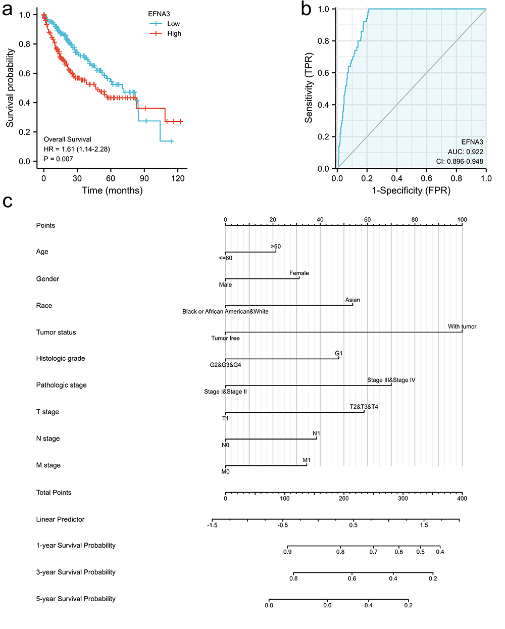
According to the survival analysis, a high level of EFNA3 expression was linked to a poor prognosis (P=0.007 in Figure 4a). High EFNA3 expression was found to be a risk factor for HCC survival in a univariate Cox regression analysis (HR=1.609, 95%CI=1.136-2.280, P=0.007), and it was discovered to be a reliable predictor of HCC survival (HR=1.606, 95%CI=1.018-2.534, P=0.042) in a multivariate Cox regression analysis (Table 2).
The area under the curve (AUC) of the ROC curve analysis of EFNA3 expression in HCC was 0.922. (Figure 4b), indicating that EFNA3 had a high diagnostic value in HCC. At the same time, in GSE14520 and GSE76427 datasets, EFNA3 had a certain diagnostic value (AUC >0.6 in Figure 5c, d). Furthermore, combining the expression of EFNA3 with clinical features, A nomogram was created to estimate the likelihood of survival for 1, 3, and 5 years (Figure 4c).
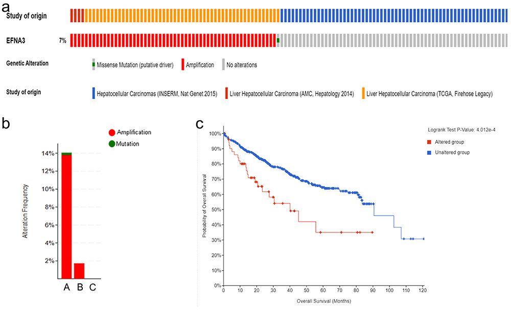
EFNA3 genetic alteration in patients with HCC
851 HCC patients in three studies (TCGA, Firehose Legacy, AMC, Hepatology 2014, INSERM, Nat Genet 2015) were analyzed on cBioPortal. In HCC, the proportion of EFNA3 alternation was 7%, and the rate of alternation was 0 to 14.06% (53/377) (Figure 6a, b). The main alternation mode is gene amplification. The KM survival curve was drawn according to the patients’ OS, the Log-rank test was conducted, and the prognosis of the population with EFNA3 gene alternation was found to be significantly worse than that of the population without EFNA3 gene alternation (P<0.05) (Figure 6c).
Immune infiltration and immune checkpoints correlation analysis of EFNA3 expression in HCC
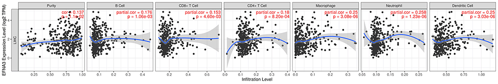
TIMER was utilized to investigate the link between EFNA3 and immune infiltration. (Figure 7). The expression of EFNA3 and the purity of HCC had a significant positive correlation. In HCC, there was also a positive association between EFNA3 expression and B cells infiltration (r=0.137, P=1.06e-03), CD4+T cells infiltration (r=0.18, P=8.20e-04), CD8+T cells infiltration (r=0.153, P=4.60e-03), Dendritic Cells infiltration (r=0.25, P=3.03e-06), neutrophils infiltration (r=0.258, P=1.23e-06). Then, in Table 3, the relationship between EFNA expression in HCC and immune cell surface markers was investigated. The expression of EFNA3 was found to be positively linked with biomarkers of B cell (CD19, CD20), T cell biomarkers (Tfh, Th1, Th2, Th9, Th17, Th22, Treg), TAM biomarker (CD80), Nature Killer cell (CD7, XCL1), Dendritic Cell (CD1C, CD11c, CD141), and Neutrophil (CD11b, CD15). But it is negatively correlated with the biomarkers of M1 macrophage (COX2, INOS) and M2 macrophage (ARG1, CD206).
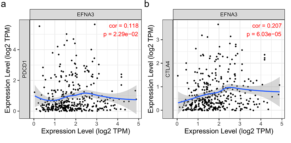
The correlation analysis between two important immunotherapy points (PDCD1, CTLA4) for HCC and the expression of EFNA3 was carried out on the TIMER website. The level of EFNA3 expression was positively correlated with PDCD1 and CTLA4 (Figure 8).
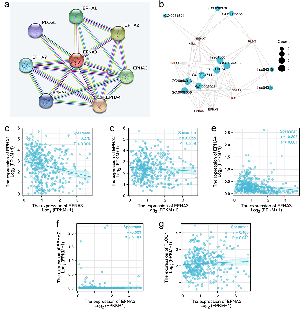
GO and KEGG enrichment analyses
Through the String database, the PPI network was formed (Figure 9a). As shown in Table 4, 7 genes highly related to the function of EFNA3 were selected (EPHA1, EPHA2, EPHA3, EPHA4, EPHA5, EPHA7, PLCG1). GO and KEGG analyses were carried out for these genes (Table 5), forming a visual network (Figure 9b). KEGG analysis found that EPHA1, EPHA2, EPHA4, EPHA7, and PLCG1 were enriched in "Axon guidance", "Rap1 signaling pathway" and "Ras signaling pathway". The expression of EFNA3 was negatively associated with EPHA1 (r=-0.270, P<0.001) and EPHA4 (r=-0.308, P<0.001), but positively correlated with PLCG1 (r=0.106, P=0.040) according to the molecular correlation analysis (Figure 9c-g).
According to GO analysis. These genes were shown to be involved in these biological processes (BP), including "ephrin receptor signaling pathway," "axon guidance," and "neuron projection guidance". "Transmembrane-ephrin receptor activity," "ephrin receptor activity," and "transmembrane receptor protein tyrosine kinase activity" were included in the molecular function (MF) level. "Schaffer collateral-CA1 synapse," "glutamatergic synapse," and "neuromuscular junction" were included in the level of cellular components (CC).
|
|
|
|
|
|
|
|
|
|
|
|
|
|
|
|
DISCUSSION |
|---|
The Eph/ephrin signaling pathway regulates Eph-interacting exchange proteins, which are involved in various related signaling pathways such as axon guidance, cell migration and apoptosis, angiogenesis and vascular smooth muscle contraction (15). EFNA3 as an Ephrin ligand has clear research significance. This study explored the expression of EFNA3 and examined the impact of EFNA3 in HCC patients.
According to the findings of this research, the EFNA3 expression level was much higher in HCC when compared with normal tissues. Combined with the correlation analysis, it showed that EFNA3 expression was positively correlated with the severity of HCC. The results also showed the expression of EFNA3 in Asians is higher than in Caucasian and black populations. This may partly explain previous conclusions regarding the highest incidence of HCC in some Asian countries (1).
Survival analysis revealed that patients with HCC who expressed a high level of EFNA3 had a worse prognosis than patients with HCC who expressed a low level of EFNA3, indicating that it might be utilized to predict the prognosis of HCC patients and EFNA3 had a good diagnostic value in HCC. Therefore, clinicians now have a nomogram to predict survival in HCC patients, which will aid doctors in forecasting patients' prognoses and making more scientific treatment decisions.
Gene alternation relates to the incidence and progression of tumors, often predicting a worse prognosis. The percentage of EFNA3 gene alternations in HCC is 7%, which was mainly gene amplification and was significantly correlated with poor OS. Gene amplification may be responsible for the high expression of EFNA3 in HCC patients.
Immunotherapy is an effective treatment for HCC. Some immune therapeutics, including therapy based on dendritic cells, immunotherapeutic checkpoint inhibitors, and adoptive cell immunotherapy, are effective for some types of HCC (16, 17). This study showed a positive association of EFNA3 with a variety of immune cells in HCC. Besides CD8+ T cells and macrophages, EFNA3 also positively correlated with the related biomarkers. Further study is needed to confirm the correlation of EFNA3 on CD8+ T cells and macrophages. Prior studies have shown that CD4+ T cells can inhibit the occurrence of HCC and mediate tumor regression (18). It has been reported that the level of T helper cell 17 (Th17) in HCC is significantly higher than that in the normal group, and it is associated with the prognosis of HCC (19). Tumor-associated macrophages (TAMs) and Regulatory T cells (Tregs) infiltration are correlated with poor prognosis in HCC (20, 21) and TAMs can contribute to the progression of HCC by promoting proliferation, angiogenesis, and subsequent metastasis (20). We found a positive correlation between EFNA3 and these cells. These findings imply that EFNA3 may reflect HCC patients’ immune microenvironment and play a key role in immune modulation. Immune checkpoint inhibitors such as PDCD1 inhibitors and CTLA4 inhibitors are considered to be effective in the treatment of HCC (6). Our results showed that EFNA3 is positively correlated with PDCD1 and CTLA4, suggesting that targeting EFNA3 could improve the efficacy of immune checkpoint inhibitors in HCC.
Molecules identified in the PPI network constructed with EFNA3 as the center were enriched in the "Axon guidance" signaling pathway, "Ras signaling pathway," and "Rap1 signaling pathway" according to KEGG enrichment analysis. In addition to axon guidance being involved in the nervous system, Ras is a membrane-associated guanine nucleotide binding protein. Actuated Ras advances cell survival and multiplication in cancer cells, and its maintenance of growth cell survival and against apoptosis are primarily mediated by PI3K/Akt and Rac/NF-κB. PI3K/Akt can deactivate B-cell lymphoma-2-associated death promoter (Bad) phosphorylation and resist apoptosis. Rac can promote actin cytoskeleton remodeling, NF-κB activation, up-regulation of B-cell lymphoma 2 (Bcl-2) and anti-apoptosis. RAS-associated protein 1 (Rap1) is a small GTPase that controls diverse processes, such as cell adhesion, cell polarity and cell-cell junction formation. It has been accounted for that Rap1b, a sub-type of Rap1 can advance the expansion, relocation and intrusion of HCC cells in vitro, as well as the event and metastasis of growths in vivo (22). EFNA3 will affect HCC through these signaling pathways.
GO enrichment analysis indicated that EFNA3 and its interacting genes were involved in “axon guidance” and “neuronal projection guidance” indicating that EFNA3 might be involved in functions of the nervous system. In addition, it is also mainly related to regulating Eph receptor activity and transmembrane receptor protein tyrosine kinase activity. These functions are correlated with HCC (7, 9), thus demonstrating the importance of EFNA3 in HCC.
This study, however, has certain limitations. The data acquired from the internet database was primarily studied in this study, and some groups were small in number, which may have led to some results not being obtained. In addition, we cannot determine the specific role of EFNA3 in the treatment of HCC due to incomplete information on clinical treatment. This still needs to be discussed in the subsequent related experiments. Presently, some researchers are conducting similar studies on Asian HCC patient datasets and applying immunochemical tests to validate the importance of EFNA3 in HCC patients (23).
|
|
CONCLUSION |
|---|
This study reveals the important clinical value of EFNA3. Meanwhile, this study found that EFNA3 is involved in immune regulation. EFNA3 was mainly involved in the Ras signaling pathway and then affected HCC. Therefore, EFNA3 can be used as a potential therapeutic target for the diagnosis or treatment of HCC.
|
|
AUTHOR CONTRIBUTIONS |
|---|
Conceptualization, methodology, formal analysis, validation and writing original draft were performed by Churun Zheng. Writing-review, editing, funding acquisition and supervision were conducted by Xiaoyu Ji and Jie Wu. All authors have read and agreed to the final version of the manuscript.
|
|
FUNDING |
|---|
This work was funded by High-level talent introduction scientific research start-up funds of Shantou University Medical College (Funding No: 510858055).
|
|
COMPETING INTEREST |
|---|
The authors mentioned that there is no conflict of interest in this study.
|
|
ETHICS APPROVAL AND CONSENT TO PARTICIPATE AND PUBLISH |
|---|
All data used in this study were open to the public. As a result, it certifies that data collection has been ethically reviewed and that written informed consent was obtained before all data collection.
|
|
REFERENCES |
|---|