| ORIGINAL ARTICLE |
|
|
1 Department of Physiology, Faculty of Basic Medical Sciences, College of Health Sciences, Ladoke Akintola University of Technology, Ogbomoso, Oyo State, Nigeria;
2 Edo University Iyamho, Iyamho, Nigeria
Corresponding Author: Serah Funke Ige, Department of Physiology, Faculty of Basic Medical Sciences, College of Health Sciences, Ladoke Akintola University of Technology, Ogbomoso, Oyo State, Nigeria. Tel: +2348060743616; E-mail: sfige@lautech.edu.ng.
FUNDING: Nil.
| |
ABSTRACT |
| INTRODUCTION | |
|
|
METHODOLOGY |
|
|
RESULT |
|
|
DISCUSSION |
|
|
CONCLUSION |
|
|
CONFLICT OF INTEREST |
|
|
ACKNOWLEDGEMENT |
|
|
REFERENCES |
|
|
ABSTRACT
|
|---|
BACKGROUND: Cadmium (Cd), a naturally occurring toxic metal, in addition to being cytotoxic, has been shown to be responsible for inducing free radical-dependent DNA damage in cells. Allium cepa is also known to ameliorate cadmium sulphate-induced organ toxicity through its antioxidant potential. The study was carried out to determine the ameliorative mechanisms of Allium cepa extract (ACE) in oxidative stress-mediated hepatic DNA damage in rats exposed to cadmium sulphate. MATERIALS AND METHODS: Twenty male adult wistar rats weighing 160-180g were used for this study. They were divided into four groups; group one (control), group two (Cd group), group three (Cd + ACE group) and group four (ACE group). Rats were administered 15 mg/kg CdSO4 and Allium cepa Extract (1 ml/100 g BW) for 28 days once per day (p.o.). Body weight changes (BWC), organ/body weight (OBW) ratio, feed conversion ratio (FCR), daily water intake (DWI) and daily feed intake (DFI) were monitored. Superoxide Dismutase (SOD), Catalase (CAT) activities and % DNA fragmentations were examined spectrophotometrically while immunohistochemical expression of Tumor Suppressor protein (p53) and cytoplasmic Bcl2 were also studied. Results were analyzed using SPSS 21 via Analysis of Variance (ANOVA). RESULTS AND DISCUSSION: Exposure to CD caused no significant change in BWC, OBW, FCR, DWI and DFI, but significantly decreased SOD and CAT activities. Significantly increase in % DNA fragmentation and Bcl2 expression and inhibited p53 expression were also observed in CD exposed rats. Decreased SOD and CAT activities, increased DNA fragmentation and Bcl2 expression together with inhibition of p53 expression orchestrated by cadmium exposure were ameliorated by Allium cepa treatments. CONCLUSION: Allium cepa remediates oxidative stress-mediated hepatic DNA damage by promoting P53 expression and inhibiting Bcl2 protein in rats exposed to cadmium sulphate.
KEY WORDS: Allium cepa; DNA fragmentation; Bcl2; Tumor suppressor protein (p53); catalase; superoxide dismutase|
|
INTRODUCTION |
|---|
Heavy metal poisoning is a form of environmental pollution bedeviling African cities (1-3). Cadmium, one of the elements of the periodic table is an example of heavy metals. It is a toxicant of environmental and industrial sources, (4). It can be found in free form and in combination with other elements, usually as cadmium chloride, cadmium nitrate and cadmium sulphate (5). Its non-degradability in the environment increases the risk of exposure and contamination (6). Human exposure to cadmium occurs through inhalation of smoke and ingestion of contaminated food and water and they are transported into body tissues where they combine with body proteins and nucleic acids eliciting a massive damage of macromolecules leading to impairment in tissue function (6). Impaired tissue function manifests as skeletal muscle injury, gastroenteritis, chronic obstructive airway disease, osteoporosis, testicular damage (1, 6, 7), renal diseases and hepatocellular damage (1).
As far as mechanistic studies on cadmium induced hepatocellular damage are concerned, modulation of tumor suppressor protein (P53) and B cell Lymphoma has been reported (6). Cytotoxic signals released by activated kupffer cells such as interleukin-1β, interleukin-6, interleukin-8 and reactive oxygen species have also been implicated (7-9). Free radicals also play role in heavy metal induced hepatic damage. Oxidants are generated by metabolism of normal cells including hepatocytes (3). However, in heavy metal induced hepatocellular damage, oxidant profile is elevated thereby instituting hepatic tissue damage. Other mechanisms of cadmium induced toxicity include deregulation of cellular response to DNA damage, resistance to apoptosis (6,10) and alteration of hepatic mitochondrial enzymes (11).
With respect to remediation of heavy metal induced hepatocellular damage, the roles of plant extracts have been documented. For instance, quercetin, a flavonoid rich extract has been shown to protect against cadmium induced hepatocellular damage by accelerating endothelial nitric oxide synthase activity (12). Ige et al., (2017) (13) also reported that extract from allium cepa mitigated aluminum chloride induced hepatic damage through regulation of oxidant/antioxidant balance. The present study hypothesized that administration of allium cepa extract has no significant effect on hepatic expressions of p53 and B cell Lymphoma in cadmium sulphate induced, oxidative stress-mediated hepatic DNA damage in rats.
|
|
METHODOLOGY |
|---|
Animal care and management
Twenty male Wistar rats weighing between 160 g-180 g were used for the study. They were obtained from the Animal house of the Department of Physiology, Ladoke Akintola University of Technology Ogbomoso, Oyo State, Nigeria. They were housed in standard cages at room temperature and 12 hr light/12 hr dark cycle. The animals were acclimatized for 1 week. All rats were fed pelletized grower mash (standard chow) and distilled water ad libitum.
Ethical certification
The study was conducted in line with the guidelines of National Institute of Health (NIH) for the use of laboratory rats.
Preparation of allium cepa (onion juice)
Fresh red onions (Allium cepa) were gotten from Waso market Ogbomoso, Oyo State, Nigeria. The onions were rinsed with distilled water and dried for 24 hours. The onions were crushed using a grating machine. Pulverization was done without addition of water. The pulverized onions were poured into a clean sieve and fluid was collected. The Allium cepa extract was prepared on daily basis as previously reported (9).
Study design
The rats were randomly divided into four groups of five rats per group.
Control group: received distill water;
Cadmium group: received daily oral administration of 15 mg/kg of CdSO4;
Cadmium + Allium cepa: received daily oral administration of 15 mg/kg of CdSO4 and 1 ml/100g of Allium cepa extract;
Allium cepa group: received daily oral administration of 1 ml/100 g of Allium cepa;
Cadmium sulphate and allium cepa extract administrations spanned for four weeks.
Tissue preparation
The rats were euthanized 24 hrs at the end of the study by cervical dislocation. The liver of the rats were excised and rinsed in 1.15% KCl. A portion of the tissue was homogenized in 0.1 mM phosphate buffer. The homogenates were centrifuged at 10,000 × g for 10 minutes and the supernatant (Post Mitochondrial Fraction) was kept under 4oC for biochemical parameters. Another portion of each tissue was homogenized in Tris-EDTA (TE) buffer and then centrifuged at 27,000 × g for 10 minutes to separate the intact DNA (pellet) from the fragmented DNA (supernatant) for DNA fragmentation. A portion of the tissue was preserved in formalin for Immunohistochemistry analysis.
Determination of hepatic antioxidant enzymes
The level of superoxide dismutase activity was determined by the method of Mistra and Fridovich (1972) (14). Catalase activity was determined according to the method Sinha (1971) (15).
Assay of DNA fragmentation by diphenylamine (DPA) method
DNA was extracted from the tissue homolysate (homogenate) and the supernatant and pellet were both subjected to diphenylamine (DPA) for colour development. The absorbance was then taken spectrophotometrically at 620nm. The percentages of genomic DNA fragmentation were then calculated.
Immunohistochemistry
A section of the liver tissue was excised and washed in 1.15% potassium chloride. The tissues were preserved in formalin for tumor suppressor protein (p53) and Bcl2 analyses. Formalin-fixed and paraffin-embedded tissue samples of the liver were cut into 4-mm sections and dried overnight at 60°C. Representative sections were stained using hematoxylin-eosin prior to immunostaining. All the slides were examined and scored independently by the observers in blinded fashion. Immunoreactivity for the tumor suppressor protein (p53) was detected using monoclonal antibody DO7 (Dako, Denmark). Tissues were considered to be positive for p53 when the cells were stained. Bcl2 expression was analyzed by using the K492 kit (Dako, Denmark). Tissues were considered to be positive when the cells accumulated cytoplasmic Bcl-2 staining.
Statistical analysis
The data obtained were expressed as mean ± standard error of mean (Mean ± SEM). Statistical analysis was done using SPSS 21. Pairwise comparison was done using least square difference. Values with p<0.05 were regarded as being significant.
|
|
RESULT |
|---|
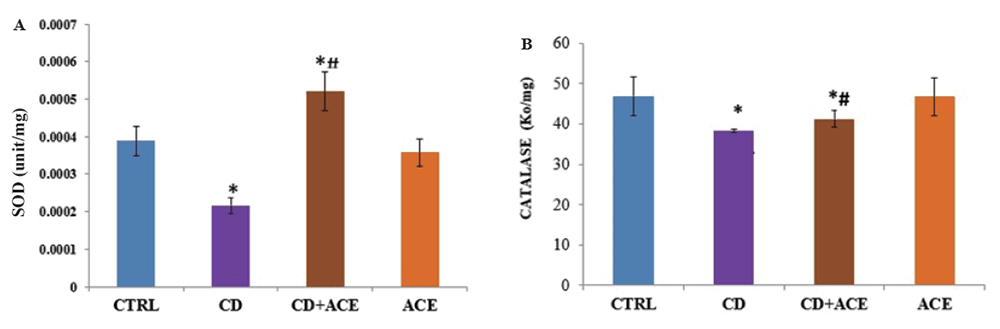
Effect of cadmium sulphate and allium cepa extract on hepatic antioxidant enzymes
Cadmium sulphate administration led to a significant decrease in SOD when compared with control. Allium cepa extract administration to cadmium sulphate treated rats led to an increase in SOD when compared with control and cadmium sulphate groups respectively (Figure 1a).
Allium cepa extract administration to cadmium sulphate treated rats led to an increase in catalase when compared with control and cadmium sulphate groups respectively (Figure 1b).
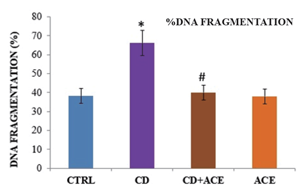
Effect of cadmium sulphate and allium cepa extract on % DNA damage
Allium cepa extract administration to cadmium sulphate treated rats led to a significant decrease in % DNA fragmentation when compared with cadmium sulphate group (Figure 2).
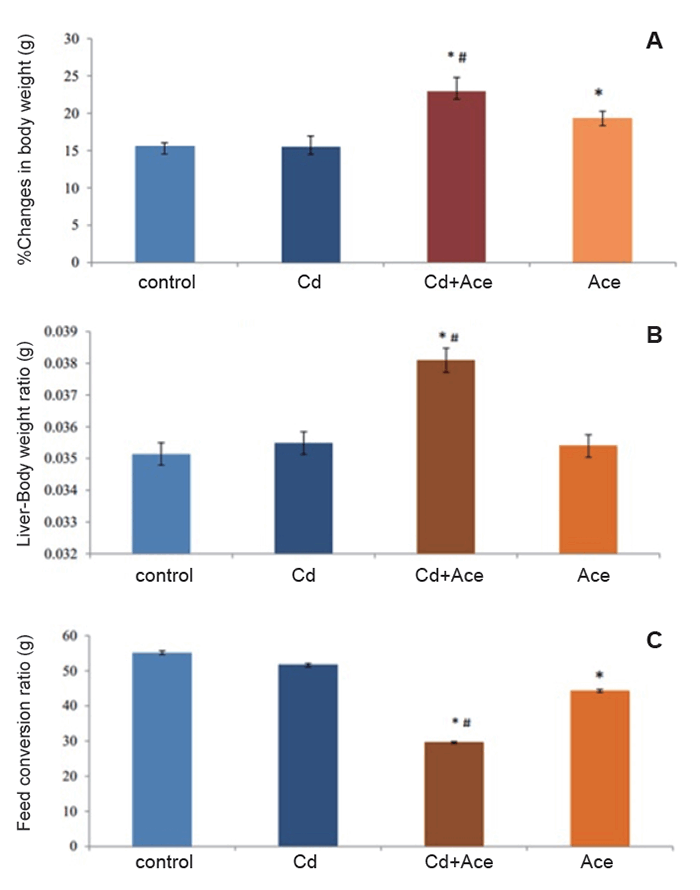
Effect of cadmium sulphate and allium cepa extract on percentage change in body weight, liver body weight ratio and feed conversion ratio.
Administration of allium cepa extract to cadmium sulphate treated rats caused a significant increase in % change in body weight of CD + ACE group when compared with the control group and cadmium group respectively (Figure 3a).
Administration of allium cepa extract to cadmium sulphate treated rats resulted in a significant increase in the liver weight change when compared with the control group and the cadmium group (Figure 3b).
Administration of allium cepa extract to cadmium sulphate treated rats caused a significant decrease in the feed conversion ratio of CD + ACE group when compared with the control group and cadmium group (Figure 3c).
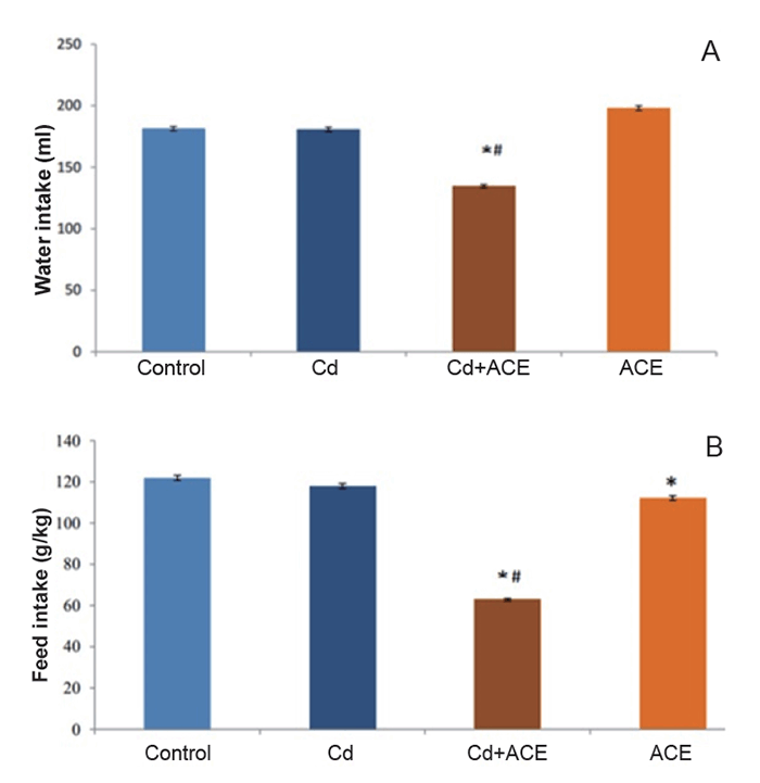
Effect of cadmium sulphate and allium cepa extract on daily water intake and daily feed intake
There was no significant difference in daily water intake of cadmium group when compared with the control group (Figure 4a). Administration of allium cepa extract to cadmium sulphate treated rats significantly decreased daily water intake when compared with control group and cadmium group respectively.
There was no significant difference in the daily feed intake of cadmium group when compared with the control group (Figure 4b). Administration of allium cepa extract to cadmium sulphate treated rats led to a significant a significant decrease in the daily feed intake when compared with the control group and cadmium group respectively.
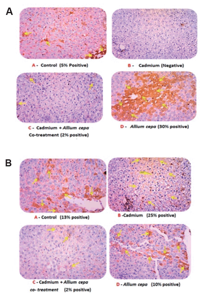
Effect of cadmium sulphate and allium cepa extract on p53 and Bcl2 expressions
Administration of allium cepa extract to cadmium sulphate treated rats culminated into mild expression of p53 expression (Figure 5a) (the yellow arrow points to region of immunoreaction). Maximum p53 expression was observed in allium cepa treated group and the least expression was in cadmium sulphate treated group.
Administration of allium cepa extract to cadmium sulphate treated rats led to relative suppression in Bcl2 expression (Figure 5b) (the yellow arrow points to region of immunoreaction). Maximum p53 expression was observed in cadmium sulphate treated group.
|
|
|
|
|
|
|
DISCUSSION |
|---|
DNA damage, impaired expressions of p53 and B cell lymphoma and disruption of oxidant/antioxidant balance have been implicated in heavy metal induced hepatocellular damage in rats. The present study investigated the ameliorative mechanisms of Allium cepa extract in oxidative stress-mediated hepatic DNA damage in rats exposed to cadmium sulphate.
The result of the study showed that exposure to cadmium sulphate led to a significant increase in % DNA damage. Previous study has shown that heavy metal exposure resulted in DNA damage through attenuation of DNA repair proteins such as nucleotide excision repair; non-homologous end joining, base excision repairs and mismatch repair (16). In addition, heavy metal induced DNA damage may also be mediated by insertion of DNA strand breaks (DSBs) to the DNA helix (16).
Another mechanism underlying heavy metal induced DNA damage is disruption of antioxidant homeostasis through increase in the production of reactive species or decrease in antioxidant capacity (7, 8,17). Like other studies (16, 18), our findings indicated that cadmium sulphate induced DNA damage was characterized by depletion in superoxide dismutase and catalase activities. Oxidants are generated by metabolism of normal cells including hepatocytes (3). However, in heavy metal induced hepatocellular damage, oxidant profile is elevated thereby instituting hepatic tissue damage.
We observed that administration of allium cepa extract to cadmium sulphate -challenged rats decreased % DNA damage when compared with rats exposed to cadmium sulphate. Previous reports on allium cepa extract indicated that it exerted a mild prevention of nephrotoxicity (9) and protected cardiac toxicity induced by exposure to cadmium respectively (18).
Tumor suppressor (p53) protein is a regulatory protein (19) that facilitates repair of DNA damage prior to replication (20, 21). Failure of DNA repair plays an important role in the carcinogenesis (21). Our immunohistochemical analysis showed that cadmium inhibits the expression of cytoplasmic tumor suppressor (p53) in the liver. Increase in hepatic expression of p53 was observed in rats treated with allium cepa extract. Administration of allium cepa extract to rats exposed to cadmium sulphate culminated into slight improvement in p53 expression.
We also observed that exposure to cadmium sulphate caused alteration in B cell lymphoma-2, an executioner protein that activates the final effector caspases of apoptosis (22). Damage to BCl-2 genes occurs in breast, lung and prostate cancers and leukemia (22). Early report by Fernandez et al., (2003) (23) has shown that cadmium exposure orchestrated an alteration in Bcl2/Bax equilibrium. In our study, we noticed that male rats treated with allium cepa extract exhibited reductive expression of Bcl2. Also, administration of allium cepa extract to rats exposed to cadmium sulphate also produced a relative reduction in Bcl2 expression.
Administration of allium cepa extract had no significant effect on hepatic superoxide and catalase when compared with control. However, we noticed significant increases in the activities of these enzymes in rats co-administered with cadmium sulphate and allium cepa extract when compared with cadmium exposed rats. This suggests that the ameliorative influence of allium cepa extract on cadmium sulphate induced hepatic DNA damage might also be mediated by a rise in hepatic antioxidant enzymes.
|
|
CONCLUSION |
|---|
The results of the study show that allium cepa extract remediates oxidative stress -mediated hepatic DNA damage through enhanced P53 expression and inhibition of Bcl2 protein in male wistar rats exposed to cadmium sulphate.
|
|
CONFLICT OF INTEREST |
|---|
The authors declare that no conflicting interests exist.
|
|
ACKNOWLEDGEMENT |
|---|
We are grateful to the technical staff of Physiology Department, Ladoke Akintola University Ogbomoso.
|
|
REFERENCES |
|---|