| ORIGINAL ARTICLE |
|
|
1 Professor and HOD, Department of Prosthodontics including Crown and Bridge and Implantology, M R Ambedkar Dental College and Hospital, Bengaluru, Karnataka, India;
2 Post-graduate student, Department of Prosthodontics including Crown and Bridge and Implantology, M R Ambedkar Dental College and Hospital, Bengaluru, Karnataka, India;
3 Reader, Department of Prosthodontics including Crown and Bridge and Implantology, M R Ambedkar Dental College and Hospital, Bengaluru, Karnataka, India;
4 Reader, Department of Prosthodontics including Crown and Bridge and Implantology, M R Ambedkar Dental College and Hospital, Bengaluru, Karnataka;
5 Post-graduate student, Department of Prosthodontics including Crown and Bridge and Implantology, M R Ambedkar Dental College and Hospital, Bengaluru, Karnataka, India;
6 Lecturer, Department of Radiodiagnosis and Imaging, D Y Patil Medical College and Research Centre, Pimpri, Pune, India
Corresponding Author: Ishita Jakhanwal, Department of Prosthodontics including Crown and Bridge and Implantology, M R Ambedkar Dental College and Hospital, Bengaluru, Karnataka, India. Tel: + 91-9535751921; E-mail: ishitajakhanwal90@gmail.com.
| |
ABSTRACT |
| INTRODUCTION | |
|
|
MATERIALS AND METHODS |
|
|
RESULTS |
|
|
DISCUSSION |
|
|
CONCLUSION |
|
|
ETHICAL CONSENT |
|
|
REFERENCES |
|
|
ABSTRACT
|
|---|
PURPOSE: To compare the marginal fit of all metal, porcelain fused to metal and all ceramic crowns fabricated using different materials and techniques commercially available. MATERIALS AND METHODS: 80 freshly extracted human mandibular first premolars were divided into 8 groups of 10 each which received Nickel-Chromium (Ni-Cr) all metal (AM) crowns, Cobalt-Chromium (Co-Cr) AM crowns, Ni-Cr three-quarter crowns, Co-Cr three quarter crowns, porcelain fused to metal (PFM) crowns with Ni-Cr copings, PFM crowns with Co-Cr copings, pressed all ceramic (AC) crowns and CAD/CAM fabricated AC crowns respectively. Crowns were cemented and specimen were sectioned buccolingually. The marginal gap was evaluated under a stereomicroscope. Lesser marginal gap indicated a better marginal fit. RESULTS: The mean marginal gap was maximum for Group 8 (222.3 μm) and least for Group 1 (85.5 μm). The mean marginal gaps for ‘all metal crowns’ and ‘metal ceramic crowns’ showed significantly lesser marginal gaps (p<0.05) for Ni-Cr Groups than Co-Cr Groups. When only ‘all ceramic crowns’ were compared, significantly lesser marginal gap was found for pressed AC crowns (148.6 μm) than CAD/CAM fabricated AC crowns (222.3 μm). CONCLUSION: Marginal fit of AM crowns were significantly better than PFM crowns and AC crowns. Ni-Cr group always showed better marginal fit than Co-Cr group. A better marginal fit of pressed AC crowns was seen than CAD/CAM fabricated AC crowns. Chamfer finish line showed a significantly better marginal fit than shoulder finish line.
KEY WORDS: Finish line; Marginal fit; Marginal gap|
|
INTRODUCTION |
|---|
Restoration can survive in the biologic environment of the oral cavity only if the margins are closely adapted to the finish line of the preparation. The configuration of the preparation finish line dictates the shape and bulk of restorative material in the margin of the restoration. It also can affect both marginal adaptation and the degree of seating of the restoration. The marginal fit of castings basically relies on perceptive tooth preparation, accurate impressions, precision castings, and careful finishing procedures (1, 2).
Precise marginal fit is essential for a successful cast restoration because intraoral degradation of cements can result in loss of marginal seal and promote retention of plaque. Thus, marginal fit of castings is one factor that leads directly or indirectly to secondary caries, adverse pulpal reactions, and periodontal disease (2).
Till this date, there is no conclusive evidence of optimum fit of crowns. This topic is heavily investigated and fit values reported are widely diverse and range from 7.5 μm to 313 μm. Such variation can be mainly attributed to the lack of coherence about the definition of “fit”, along with differences in methods employed to determine the fit, testing parameters followed and various materials used for the fabrication of crowns (3, 4).
This study was done to compare and evaluate the marginal fit of complete metal, porcelain fused to metal and metal free ceramic crowns fabricated with different types of materials commercially available, in which crowns were fabricated for mandibular first premolars and cemented using resin cement. The cement thickness at the margins were measured using stereomicroscope. This study thus aimed to determine the best marginal fit amongst the mentioned crowns above.
|
|
MATERIALS AND METHODS |
|---|
Human first mandibular premolars, eighty in number, extracted for orthodontic purposes or periodontally compromised teeth (indicated for extraction) were selected using the following inclusion criteria:
Completely formed apex.
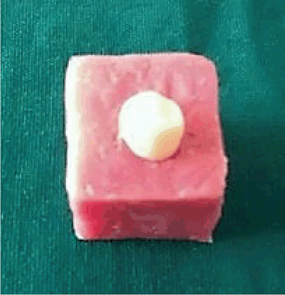
After removal of calculus and soft tissue deposits, teeth were disinfected in 3% sodium hypochlorite solution and embedded in standard MS (mild steel) cylinders (20 mm diameter, 20 mm length) with self curing acrylic resin (DPI, India) to within 2 mm of the cemento-enamel junction, with the long axis of the roots perpendicular to the horizontal plane (Fig. 1). They were divided into eight groups of ten each to receive the following groups of crowns: Group 1 - Nickel-Chromium (Ni-Cr) all metal (AM) crowns; Group 2 - Cobalt-Chromium (Co-Cr) AM crowns; Group 3 - Ni-Cr three-quarter crowns; Group 4 - Co-Cr three quarter crowns; Group 5 - porcelain fused to metal (PFM) crowns with Ni-Cr copings; Group 6 - PFM crowns with Co-Cr copings; Group 7 - pressed all ceramic (AC) crowns and Group 8 - CAD/CAM fabricated AC crowns.
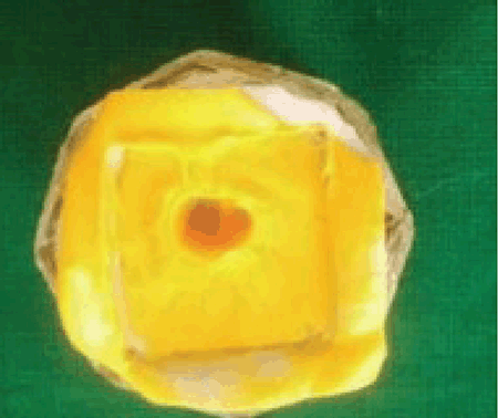
Ideal tooth preparation was done for each group to receive ‘all metal, porcelain fused to metal and all ceramic’ crowns. Group 1, 2, 3 and 4 were given chamfer finish line all around, group 5 and 6 received buccal shoulder and lingual chamfer finish lines while group 7 and 8 were prepared with all around shoulder. Impression was made for each specimen using polyvinyl siloxane (PVS) (Fig. 2) and impressions were poured in type IV die stone.
Crown fabrication for Group 1, 2, 3 and 4
Two layers of die spacer was applied and wax patterns of full coverage restoration were made for Group 1 and 2. Group 1 specimen were casted in Ni-Cr alloy (Bego, Germany), whereas Group 2 specimen were casted in Co-Cr alloy (Bego, Germany). Similarly, wax patterns for three-quarter crowns were made for Group 3 and 4 and Group 3 patterns were casted in Ni-Cr alloy, whereas Group 4 patterns were casted in Co-Cr alloy.
Crown fabrication for Group 5 and 6
Two layers of die spacer was applied and full coverage wax patterns were made for Group 5 and 6. ‘Cut Back’ of the wax patterns were done for uniform layering of the porcelain. Wax patterns were then casted in Ni-Cr alloy for Group 5 and in Co-Cr alloy for Group 6, thereby fabricating the respective metal copings. Layering of the porcelain (VITA VMK Master, Germany) was done, followed by firing (at 650°C for 15 minutes) and glazing according to the manufacturer’s instructions.
Crown fabrication for Group 7
Two layers of die spacers were applied over the dies and wax patterns with a cut back design were prepared for Group 7. Burn out of the wax patterns were done after which the investment ring was transferred to the press machine for the press procedure. The e-max glass ceramic ingots (IPS e.max, Ivoclar Vivadent, India) placed in the machine were softened at 920°C and were automatically pressed into the mould. After pressing and cooling, crowns were divested, cleaned, dried and abraded with 50 μm glass bead particles for 20 sec. Ceramic layering was done over the copings and then fired at 750°C. This was followed by glazing at a temperature of 730°C.
Crown fabrication for Group 8
The restoration for Group 8 were fabricated using computer aided designing (CAD) and computer aided machining (CAM) by CERAMILL system. Dies prepared for the Group 6 were scanned and the digital models were constructed from them. Spacer thickness of 50 μm was adjusted in the system. Virtual designing of the coping was then done in the CAD software of CERAMILL. After completion of designing, data was then fed into the milling unit to get the corresponding zirconium oxide copings (Amann Girrbach). Copings obtained were then sintered at 1450°C for 10 hours. Ceramic layering was done over the copings and then fired at 930°C. This was followed by glazing at 910°C.
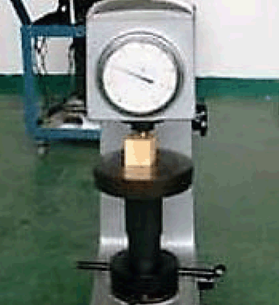
The crowns after fabrication were cemented to their respective dies with the rein cement (Maxcem Elite, Kerr Co., USA). Pink colour was added to the cement before cementation for easy visualization under the microscope. A thin, even layer of the mixed cements was applied to the fitting surface of the crowns. The crowns were seated on their respective restorations and an uniform load of 50N was applied on each crown for 2-3 minutes (Fig. 3). Final curing was done using ultra curing light for 10 sec. Curing was as per manufacturer’s instructions. Excess cement was removed from around the margins using a sharp probe.
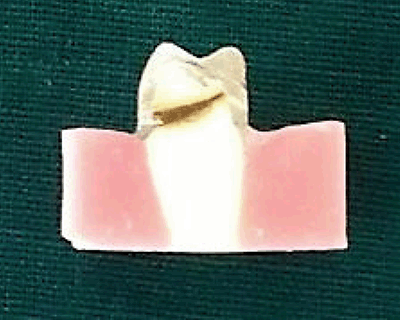
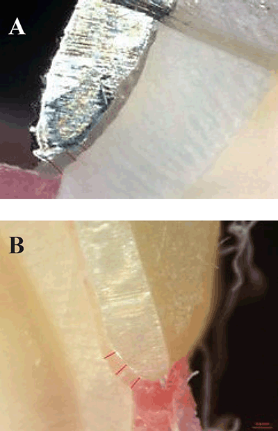
The cemented crowns were sectioned using a low speed diamond cutter (Model: Struers: Minitom) buccolingually (Fig. 4). The marginal fit was evaluated by measuring the perpendicular distance from the internal surface of the casting to the axial wall of the prepared tooth at the marginin a stereomicroscope (Model:Carl Zeiss – Discovery V20) under 40 × magnification, with an imaging processing software (Fig. 5A, 5B). 6 readings were taken for each specimen, 3 on the buccal margin and 3 on the lingual margin. The first reading was recorded at the edge of the buccal margin of the prepared tooth. The second and the third readings were made at a distance of 0.5 mm and 1mm from the point of the first reading respectively. The 3 readings were then averaged to obtain the buccal marginal gap or discrepancy. 3 readings were taken on the lingual margin in a similar way and they were averaged to obtain the lingual marginal gap or discrepancy for each specimen.
Statistical Analysis
To compare the marginal fit between the six groups, obtained results were statistically analyzed using SPSS software. The data were described in Mean, Standard Deviation and Range values. One way ANOVA was used for multiple group comparison followed by Independent sample t test to assess any significant difference between the buccal and lingual marginal gap within each group. In order to find out which pair of groups there existed a significant difference, Tukey test (post hoc test) was applied.
|
|
|
|
|
|
|
RESULTS |
|---|
ANOVA test was done to compare the mean marginal gap amongst different groups. Highest mean marginal gap was obtained for Group 8 (222.3 μm) followed by Group 6 (194.4 μm), Group 7 (148.6 μm), Group 5 (143.9 μm), Group 4 (123.4 μm), Group 2 (117.1 μm), Group 3 (94.15 μm) and Group 1 (85.5 μm) (Table 1, Table 2). In order to find out which pair of groups there exist a significant difference, Tukey test (post hoc test) was applied.
When the mean marginal gaps of ‘all metal crowns’ were compared, results showed that Group 1 showed significantly lesser value than Group 2 (P<0.05). Similarly, Group 3 showed significantly lesser marginal gap compared to Group 4 (P<0.05). However, the mean marginal gap of Group 1 showed no significant difference compared to Group 3 (P>0.05). Similarly, the mean marginal gap of Group 2 was comparable with that of Group 4 (P>0.05) (Table 3).
Comparing the mean marginal gap between the two types of ‘all ceramic crowns’, the results showed a significantly lesser marginal gap in Group 7 than Group 8 (P<0.05). When the mean marginal gap of ‘metal ceramic crowns’ were compared with ‘all metal crowns’ having similar alloy types, Group 1 showed a significantly lesser marginal gap as compared to Group 5 (P<0.05). Similarly, Group 2 showed a significantly lesser marginal gap compared to Group 6 (P<0.05) (Table 4).
When comparing the different ‘all metal crowns’ with ‘all ceramic crowns’, all the all metal groups (Group 1, Group 2, Group 3, Group 4) showed a significantly lesser mean marginal gap compared to the all ceramic crowns groups (Groups 7, Group 8) (P<0.05) (Table 5).
When the different ‘metal ceramic crowns’ were compared with ‘all ceramic crowns’, there was no statistically significant difference in the mean marginal gap of Group 5 and Group 7 (P>0.05), however, Group 5 showed a significantly lesser marginal gap compared to Group 8 (P<0.05). Furthermore, results showed that the mean marginal gap of Group 6 was significantly lesser than Group 7 and group 8 (P<0.05) (Table 6).
To find out which group exhibited significant difference between its buccal and the lingual marginal fit, Independent 2 samples ‘t’ test was performed. When the mean marginal gap between the buccal and the lingual margins for each group was compared; Group 1, Group 2, Group 3, Group 4, group 6 and Group 8 showed no significant difference for either of the margins (P>0.05). However, for Group 5 and Group 6, the mean lingual marginal gap was significantly lesser than the mean buccal marginal gap (P<0.05).
|
|
|
|
|
|
|
|
DISCUSSION |
|---|
Marginal adaptation is one of the most important criteria for determining the clinical success of the dental restoration. Poor fit of the crown can lead to deficient margins, and this could lead to tooth sensitivity in vital teeth and a higher plaque index and thereby causing caries and inflammation of periodontal tissues (5).
A definition of marginal fit varies from one study to another, with each study drawing a conclusion based on its own definition. In this study, we defined a perpendicular distance from the internal surface of the casting to the axial wall of the prepared tooth at the margin as the marginal discrepancy or the marginal gap, that can be measured only on sections of cemented crowns. In the cross-sectional technique used in the present study, the samples must be sacrificed and also it is more time-consuming. However, it has greater precision in measurements, as measurement points are more accurate and repeatable. Therefore this method was chosen for measuring the marginal fit (6, 7).
Human mandibular first premolars extracted for periodontal and orthodontic reasons were used as the test specimens for this study. The use of natural teeth is clinically more relevant compared to alternative test specimens of metal dies of typhodont teeth that have been reported on previously. The true effect on the marginal fit of the crowns may be compromised in the studies that do not use natural teeth (8).
The data supports the rejection of the null hypothesis that the marginal fit of all metal, porcelain fused to metal and all ceramic crowns are not significantly different (P<0.05). The results show that the mean marginal gap was largest for Group 8, i.e, Amann Girrbach all ceramic crowns (222.3 μm), whereas the least marginal gap was seen for group 1, i.e, Ni-Cr all metal crowns (85.5 μm). Amongst the all metal crowns, the mean marginal gap for Ni-Cr full coverage restorations were significantly lesser (85.5μm) than for the Co-Cr full coverage restorations (117.1 μm). A similar study was done by Katta Sridhar Chowdhary, in 2011, who concluded that Co-Cr alloy copings had lesser marginal fit when compared with copings of Ni-Cr alloy. The reason for the higher marginal discrepancy for Co-Cr crowns can be attributed to its higher casting shrinkage (2.3%) than that of Ni-Cr crowns (2%). This in turn may be a result of the higher melting temperature of Co-Cr alloy (1495°C) than Ni-Cr alloy (1400°C) (9).
This study also reported a significantly higher marginal discrepancy of the porcelain fused to metal crowns than the corresponding all metal crowns. The reason for the increased marginal gap after veneering was due to the difference in thermal contraction between the metal coping and the layering porcelain during cooling of the prosthesis from the firing temperature. The distortion of the lower metal core occured due to the difference in heat expansion coefficients of the lower metal and the upper porcelain, difference in the degree of expansion and high temperature during the firing of the upper porcelain. Similar findings were reported by Buchanan et al (10).
All-ceramic restorations are in fast progression in properties and usage. In situations with high esthetic demands, these materials may be the material of choice for the restoration (11). In the present study, the mean marginal gap of IPS e max, pressed glass ceramic crowns was 148.6 μm and for CAD/CAM fabricated zirconia crowns, it was 222.3 μm. Low shrinkage of heat pressed cores may explain this difference. Furthermore, in heat pressed ceramics, which uses lost wax technique, 0.4% shrinkage of wax pattern and 0.2% shrinkage of ceramic coping can be compensated by 0.3% setting and 0.2% thermal expansion of investment material. Thus, because of inherent properties, large gaps are not expected in the pressed copings. Similar results were noted by Farid et al. in their study (12). This also explains the reason for the CAD/CAM fabricated zirconia crowns having the largest marginal discrepancy when compared to the other seven groups, as all other groups except group 8 used the lost wax technique for the fabrication of the crowns. Moreover, CAD/CAM machining of the pre-sintered Y-TZP blocks induce microcracks at zirconia surface which cannot be completely eliminated by sintering. These microcracks act as stress concentration sites which together with those caused by (core veneer) CTEs mismatch are thought to contribute to the marginal distortion developed during veneering of CAD/CAM frameworks (13).
Previous studies state the amount distortion in the pressed ceramic copings is less than that observed in the metal copings. The pressed ceramic crowns have a better adaptation than the metal ceramic crowns even after exposure to multiple high temperature firing cycles (7). However, no statistically significant difference between the marginal fit of porcelain fused to metal crowns with Ni-Cr metal copings (146.7 μm) and those of pressed all ceramic crowns (IPS e max) (148.6 μm) in the present study could be attributed to the difference in the margin designs for the two types of crowns. In this study, the specimen which received porcelain fused to metal crowns with Ni-Cr copings were prepared with lingual chamfer and buccal shoulder finish line margin design, whereas specimen for the pressed all ceramic crowns (IPS e max) were prepared with chamfer finish line margin design all around. Porcelain firing cycles change the marginal fit of shoulder copings more adversely compared with rounded chamfer copings. This phenomenon occurs because the chamfer finish line has some length on axial wall of the preparation, so the closing of margin is more probable along this length. On the other hand, shoulder margin has a butt joint form, without any length on the axial wall. This is why if any distortion happens due to porcelain firing, it will affect the whole marginal gap. Pera et al., evaluated the marginal adaptation of porcelain ceramic crowns and reported improved marginal fit of In-Ceram crowns fabricated on chamfer compared with shoulder finish line (14).
The result of the present study was in accordance with the aforesaid studies, explaining the reason for a statistically significant difference between the marginal discrepancies of the buccal and the lingual margins in the porcelain fused to metal group. Similary, no significant difference was found between the buccal and lingual marginal gaps within group 7 and group 8 as they exhibited shoulder margin design all around.
|
|
CONCLUSION |
|---|
Moldovan et al. in his study, rated the values of 100 μm for marginal misfit as ‘good’ and values of 200–300 μm as ‘acceptable’ (7). According to the above, the marginal misfit or discrepancy for the ‘all metal’ crowns noted in the present study was within 100 μm and thus, could be rated as ‘good’. For the ‘porcelain-fused-to-metal’ crowns and ‘all ceramic’ crowns, the marginal misfit or discrepancy obtained was between 200-300 μm which could be rated as ‘acceptable’.
Within the limitations of the study, the marginal fit of all the types of crowns being within the clinically acceptable range, it was concluded that the marginal fit of ‘all metal crowns’ were significantly better than ‘porcelain fused to metal’ or ‘all ceramic restorations’. Moreover, Ni-Cr crowns had a significantly better marginal fit than Co-Cr crowns. Porcelain fused to metal crowns showed a better marginal fit with Ni-Cr copings as compared to Co-Cr copings. The study also concluded a better marginal fit of pressed all ceramic crowns than CAD/CAM fabricated all ceramic crowns. When compared to porcelain fused to metal crowns, pressed all ceramic crowns showed almost comparable marginal fit with metal ceramic crowns with Ni-Cr copings, however a better marginal fit than metal ceramic crowns with Co-Cr copings. No significant difference was found between the full coverage and partial coverage all metal restorations. Chamfer finish line design showed a significantly better marginal fit than shoulder finish line design.
|
|
ETHICAL CONSENT |
|---|
The present study was performed according to the ethical guidelines and was approved by the Institutional Ethics Committee. The study does not involve human beings. It was an ‘In vitro’ study performed on first mandibular premolars, extracted for orthodontic purposes or periodontally compromised teeth (indicated for extraction), having the inclusion criteria already mentioned. No healthy teeth were extracted.
|
|
REFERENCES |
|---|