| ORIGINAL ARTICLE |
|
|
1 Advanced Magnetic Resonance Center, Oklahoma Medical Research Foundation, 825 NE 13th street, Oklahoma City, Oklahoma, USA;
2 Department of Pharmaceutical Sciences, The University of Oklahoma College of Pharmacy, 1110 N. Stonewall Avenue, Oklahoma City, Oklahoma, USA
Corresponding Author: Hariprasad Gali, The University of Oklahoma College of Pharmacy, 1110 N. Stonewall Avenue, Room 301, Oklahoma City, OK 73117, USA. Tel: (405) 271-6593 Ext 47877; Fax: (405) 271-7505; E-mail: hgali@ouhsc.edu.
Running title: Hydroxymethylphosphine oxide detection in vivo by 31P-MRS
| |
ABSTRACT |
| BACKGROUND | |
|
|
METHODS |
|
|
31P MRS |
|
|
RESULTS AND DISCUSSION |
|
|
ACKNOWLEDGEMENTS |
|
|
ABBREVATIONS |
|
|
CONFLICT OF INTEREST |
|
|
REFERENCES |
|
|
ABSTRACT
|
|---|
Application of organophosphorus compounds in biomedicine is attractive because the 31P nucleus is very amenable to study by nuclear magnetic resonance (NMR) techniques, particularly, by in vivo 31P magnetic resonance spectroscopy (31P-MRS). The water-soluble organophosphorus compounds that are non-toxic, exhibit metabolic stability, and show a unique resonance peak in 31P NMR spectroscopy, which could be ideal to be used as probes for 31P-MRS. Here we evaluated the in vivo feasibility of potentially using a hydroxymethylphosphine oxide as a novel probe for 31P-MRS studies using tris(hydroxymethyl)phosphine oxide (THPO) as an example. THPO was synthesized, injected in the normal CF1 mice, and 31P spectra were acquired before and after injection with the coil located on the regions of interest. The NMR signal from the region of interest appeared within one minute of THPO injection. The compound was stable in vivo as no metabolites of THPO were observed. No toxicity was observed after THPO injection in mice. The peak concentrations of THPO in liver and kidney were reached within 15 min and 60 min respectively. THPO was excreted exclusively in urine without undergoing any metabolism indicating that it is very stable under in vivo conditions. These initial studies in normal CF1 mice clearly demonstrate that THPO possess the essential characteristics required for a potential MRS probe. Based on the current preliminary results, we suggest that HMPs, when incorporated into targeted drugs (peptides, proteins, antibodies, etc.), may serve as novel 31P probes for monitoring the drug distribution in vivo by MRS.
KEY WORDS: 31P-MRS; hydroxymethylphosphine oxide; MRI; NMR|
|
BACKGROUND |
|---|
Application of organophosphorus compounds in biomedicine is attractive because the 31P nucleus is very amenable to study by nuclear magnetic resonance (NMR) techniques, particularly, by in vivo 31P magnetic resonance spectroscopy (31P-MRS). The advantages of using 31P-MRS are: a) 31P is a desirable NMR nucleus with 100% natural abundance, high NMR sensitivity (~6.6% of 1H), relatively large g value (10,830 rad/gauss), and a large chemical shift range (1); b) the technique is intrinsically noninvasive; c) there is no radioactivity involved and therefore the compounds are inherently stable with a long shelf life, allowing studies at later time points after injection when the blood background is diminished; and d) it is possible to obtain high-resolution anatomic 1H magnetic resonance imaging (MRI) in conjunction with the 31P NMR spectra, which provide context in terms of organs and tissue heterogeneity, with no need for imaging after processing and co-registration procedures.
The organophosphorus compounds that are non-toxic, soluble in biologically compatible solvents, exhibit metabolic stability, and show a unique resonance peak in 31P NMR spectroscopy, are ideal to be used as probes for 31P-MRS. Here we discuss the utility of a tris(hydroxymethyl)phosphine oxide (THPO) as a probe for 31P MRS. Little is known regarding the toxicity of THPO. THPO was not found to be mutagenic in Salmonella and did not inhibit acetyl cholinesterase in vitro (2, 3). THPO was nominated by the National Toxicology Program in 2005 to undergo toxicity tests, since it is a by-product and metabolite of tetrakis(hydroxymethyl)phosphonium chloride (THPC), a reactive flame retardant widely used with cellulosic and cellulosic blend fabrics, including FR-treated infant sleepwear and work clothes (4).
Katti et al. have reported the utility of hydroxymethylphosphines as chelating ligands for labeling biomolecules with 99mTc/188Re for developing targeted diagnostic/therapeutic radiopharmaceuticals (5-7). However, no organophosphorus compound has ever been utilized for any other diagnostic application. To our knowledge, this is the first report that describes the utility of an organophosphorus compound as an extrinsic 31P-MRS probe. 31P-MRS is currently used to determine intrinsic phosphorus metabolite concentrations in human tissues such as muscle and brain (8-9).
|
|
METHODS |
|---|
Materials
Tetrakis(hydroxymethyl)phosphonium chloride, triethylamine, and 30 wt% hydrogen peroxide were purchased from Sigma-Aldrich (St. Louis, MO). Tris(hydroxymethyl)phosphine (THP) was synthesized by slight modifications to the previously reported procedure (10). 31P NMR spectroscopy was performed on a Varian Mercury VX-300 NMR Spectrometer at the NMR Facility (University of Oklahoma, Norman, OK). CF-1 (19-21 g) male mice were purchased from Charles River Laboratories International, Inc. (Wilmington, MA). All animal studies were conducted in accordance with the protocols approved by both the OUHSC and OMRF institutional animal care and use committees. All MR experiments were conducted using a 2.0 cm surface coil in a 7 Tesla 30 cm horizontal bore small animal MRI system (Bruker BioSpin MRI, Germany) at the Advanced Magnetic Resonance Center at the Oklahoma Medical Research Foundation, Oklahoma City, OK.
Tetrakis (hydroxymethyl) phosphine oxide (THPO)
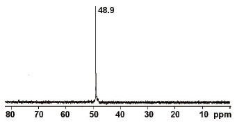
To a solution of THP (3.4 g, 27.4 mmol) in water (6 ml) was added 30 wt% hydrogen peroxide in water (8.8 ml, 77.6 mmol). The reaction mixture was heated to 35 0C for 30 min. The progress of the reaction was monitored by 31P NMR. After completion of the reaction, the water was evaporated on rotary evaporator to obtain the THPO as a white solid (yield - 3.6 g, 93.8%). The THPO was used without any further purification. 1P NMR (D2O, 121.47 MHz) δ (ppm) 48.9 (s, 1P); 13C NMR (D2O, 75.45 MHz) δ (ppm) 52.2 (d, 3C).
|
|
31P MRS |
|---|
THPO frequency
To determine the frequency of the THPO, two 1.5 ml centrifuge tubes (one containing 1M phosphoric acid as a reference and the other containing 1M THPO solution) were placed at the isocenter of the magnet and a 31P single pulse spectrum was acquired with a repetition time (TR) of 3,000 ms, 2,048 points, an acquisition time of 146.23 ms and a spectral width of 14 kHz. The THPO frequency was determined to be 121.589 MHz.
T1 relaxation time of THPO
To identify the T1 relaxation time of the THPO, a series of 31P RARE images with varying TR were acquired on a 1.5 ml centrifuge tube containing 1M THPO solution (scan parameters: echo time (TE) = 14.4 ms; TR = 250, 500, 750, 1,000, 1,500, 2,000, 2,500, 3,000, 4,000 and 5,000 ms; matrix size = 32 × 32 pixels; field of view = 8 × 8 cm2; 1 axial slice of 5 mm thickness). Regions of interest were drawn on the tube to get signal intensity measurements that were fitted to an equation linking signal intensity to TR:
|
The T1 of THPO was determined to be 2,990 ms. As a consequence, a TR of 3,000 ms was chosen for subsequent experiments.
Animal experiments
Animal experiments were conducted at a frequency of 121.589 MHz using a 31P single pulse sequence over a spectral width of 8,000 Hz, 32 averages, a TR of 3,000 ms, 2,048 points, and an acquisition time of 1 min 36 s. A 72 mm-diameter 1H/31P resonator was used for signal transmission and the signal was received through a tunable 1H/31P flat surface coil positioned on the region of interest of the sample (phantom or mouse). Normal CF-1 mice were anesthetized initially using 2% isoflurane in oxygen at 2 L/min, in a polypropylene induction chamber. When fully anesthetized, a catheter was placed in the tail vein and the animals were placed on the scanner bed. Anesthesia was maintained through a nose cone with 1% isoflurane in oxygen at 2 L/min. Body temperature was maintained at 37 ± 1°C by using a water-circulated pad under the animal. Animals were placed in a supine position for the liver experiments, or on its side for the kidney experiments, and the 1H/31P surface coil was located on the organs of interest. A quick 1H-T1-weighted morphological image was acquired to guarantee the proper positioning of the surface coil, and a 31P “baseline” spectrum was acquired. 100 µl of 4M THPO solution was then administered through the tail vein, and 31P spectrum acquisition was resumed for 2 hours (liver experiment) or 4 hours (kidney experiment). Following MR experiments, the mice were euthanized, urine and blood of the animal were collected, and 31P spectra were acquired on these ex vivo samples. 31P spectra were pre-processed using the Bruker TopSpin software (apodization, Fourier transformation, and phasing of spectra), and then calibrated and integrated to provide peak areas using Mathematica (Wolfram Research).
|
|
RESULTS AND DISCUSSION |
|---|
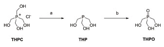
To demonstrate the proof-of-concept, we synthesized THPO and evaluated the feasibility of observing this compound using 31P-MRS in normal CF1 mice. THPO was synthesized in a two-step reaction as shown in Figure 1. First, commercially available THPC was treated with triethylamine to obtain THP and then it was oxidized with hydrogen peroxide to obtain THPO. THPO is soluble in water at all concentrations due to the presence of hydrophilic -CH2OH substituents, and presents a single peak in 31P NMR spectra with a chemical shift of ~49 ppm as shown in Figure 2. This chemical shift is farther down field than that of all indigenous phosphorous containing compounds (<20 ppm) found in the body.
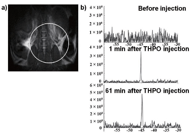
For in vivo evaluation, we injected THPO solution in the first mouse and acquired 31P spectra before and after injection with the coil located between the kidneys and the bladder, as shown in Figure 3. The NMR signal from the region covering the kidneys and the bladder appeared within one minute of injection. The compound was stable in vivo as no metabolites of THPO were observed. Only a single peak corresponding to THPO was observed throughout the imaging period of 61 min (Figure 3b). After THPO injection, the mouse behaved normally and no adverse reactions were observed.
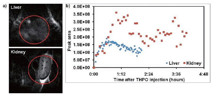
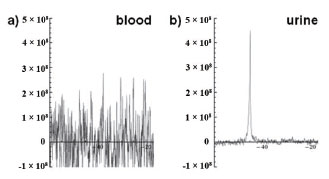
To follow the clearance of THPO from the blood pool, we injected two mice with THPO and assessed the liver in one mouse for 2 h, and the kidney in the other mouse for 4 h (Figure 4). The mice were killed, and the blood and urine were collected at the end of the imaging study. As shown in the Figure 4b, the peak concentrations in the liver and the kidneys were reached within 15 min and 60 min, respectively. Since THPO is very hydrophilic, as expected it was cleared predominantly through the kidneys into urine (Figure 5). The clearance from the kidneys was relatively slower because THPO is a small hydrophilic and neutral molecule that may be just cleared by glomerular filtration but not secreted by the tubules. As shown in Figure 5a, no THPO was observed in the blood samples collected from the mice indicating that THPO was completely cleared from the blood pool by 2 h. THPO was excreted exclusively in urine without undergoing any metabolism indicating that it is very stable under in vivo conditions (Figure 5b). More in vivo studies are needed to determine the pharmacokinetic properties of THPO, as well as toxicity. These initial studies in normal CF1 mice clearly demonstrate that THPO possess the essential characteristics required for a potential MRS probe.
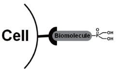
New HMPO derivatives containing functional groups such as –COOH that allow conjugation to biomolecules (e.g., peptides, proteins, antibodies, etc.), as shown in Figure 6, are needed to increase the concentration of the probe at the targeted disease site. The hydrophilic nature of HMPOs facilitates their efficient clearance from the blood pool primarily through the renal/urinary pathway if they are separated from biomolecules due to any in vivo degradation process. Thus, this minimizes the background signal due to presence of free HMPO in the blood pool. However, the biodistribution and pharmacokinetic properties of the HMPO-biomolecule conjugate may predominantly be dominated by those of the biomolecule itself.
Based on the current preliminary results, we suggest that HMPOs, when incorporated into targeted drugs (peptides, proteins, antibodies, etc.), may serve as novel 31P probes for monitoring the drug distribution in vivo by MRS. However, due to the inherent low sensitivity, it is a challenging endeavor to develop a molecular-targeting MRS probe. We will undertake further studies to address the critical issue of sensitivity with HMPO based molecular-targeting MRS probes in the future.
|
|
|
|
|
|
|
|
ACKNOWLEDGEMENTS |
|---|
This work was funded by the University of Oklahoma College of Pharmacy Startup Grant. We gratefully acknowledge the expert technical assistance of Debra Saunders during animal studies.
|
|
ABBREVATIONS |
|---|
HMPO, Hydroxymethylphosphine oxide; THPO, Tris (hydroxymethyl) phosphine oxide; THPC, Tetrakis (hydroxymethyl) phosphonium chloride; THP, Tris (hydroxymethyl) phosphine; MRS, Magnetic resonance spectroscopy; MR, Magnetic resonance; NMR, Nuclear magnetic resonance; ppm, Parts per million; TR, Repetition time; TE, Echo time.
|
|
CONFLICT OF INTEREST |
|---|
The authors declare that no conflicting interests exist.
|
|
REFERENCES |
|---|