| ORIGINAL ARTICLE |
|
|
1 Diabetes and Endocrine Unit, Cardiovascular, Diabetes and Nutrition Research Centre (CDNRC);
2 Toxicology and Pharmacology Unit, Herbal Medicine Research Centre (HMRC);
3 Phytochemistry Unit, Herbal Medicine Research Centre (HMRC), Institute for Medical Research, Jalan Pahang, 50588, Kuala Lumpur, Malaysia
Corresponding Author: Ezarul Faradianna, Diabetes and Endocrine Unit, CDNRC, Malaysia. E-mail: fara@imr.gov.my.
| |
ABSTRACT |
| INTRODUCTION | |
|
|
MATERIALS AND METHODS |
|
|
RESULTS |
|
|
DISCUSSION |
|
|
CONCLUSIONS |
|
|
ACKNOWLEDGEMENT |
|
|
CONFLICT OF INTEREST |
|
|
REFERENCES |
|
|
ABSTRACT
|
|---|
Dioscorea hispida (D.hispida) is the most well-known starchy tuber in Malaysia and called ‘ubi gadong’. Despite concerns over toxicity effects, the tuber is known to possess therapeutic values due to the presence of bioactive compounds such as saponins. This study was performed to identify the changes in gene expression profiles associated with hepatoxicity in pregnant rat treated with D.hispida using RT² Profiler PCR Array. The identification of steroidal saponins from D.hispida was carried out by UHPLC/MS method. Treatment of D.hispida caused mortality when dosage above 2000 mg/kg b.w. was given to pregnant rats. The PCR array showed that several genes were significantly up and down-regulated upon treatment with D.hispida. Treatment of D.hispida at 2000 mg/kg b.w leads to significant upregulation of several genes such as Btg2, Gsr, L2hgdn, S100a8, Slc17a3, Bhmt, Cd68, Cyp1a2 whereas several genes were downregulated such as Abcb1a, Aldoa, Cdc14b, Icam1, Krt18, Hpn and Maob. The consumption of D.hispida extract when taken at lower dosage of 2000 mg/kg may not be harmful to rats. D.hispida extract given at the highest dosage to pregnant rats caused alterations of several genes categorized in different hepatotoxic group functions such as necrosis, cholestasis and phospholipodisis.
KEY WORDS: hepatoxicity; Dioscorea hispida; pregnant rat; gene expression|
|
INTRODUCTION |
|---|
Dioscorea (Family Dioscoreacea) commonly known as yams, are staple foods for Africa, tropical America and Asia. Yams have been traditional consumed as starchy staple food (such as Dioscorea opposita, Dioscorea alata, Dioscorea japonica, and Dioscorea hispida). The genus Dioscorea L. consists of about 630 species distributed all over the world. Dioscorea hispida is the most well-known starchy tuber in Malaysia and it is called `ubi gadong’. Majority of this species contain steroidal saponins with potent pharmacologic activities. Some of Dioscorea species being the source of the steroidal sapogenin, diosgenin a precursor for the synthesis of steroidal drugs. The glycoside form of diosgenin is called Dioscin (1).
Many of the yam tubers are reported to be toxic containing cyanides and have to be detoxified by soaking in flowing water and cooking before consumption in order to remove the toxic compound presents in them (2, 3). However, since traditional detoxified methods are difficult to practice, new dioscorine detoxified equipment was developed to improve the quality and time of detoxified process (4, 5). In terms of therapeutic values, several pharmacological studies have demonstrated that Dioscin exhibits anti-tumor (6), anti-hyperlipidemia (7), and anti-diabetic (8) properties. In addition, D. hispida extract exhibited significant anti-inflammatory and analgesic properties (9). The protective effect of D. alata however has also been shown previously in aniline-induced splenic toxicity rats due to its antioxidant property and the presence of different phytochemicals (10). D. alata contained alkaloid, flavanoid, saponin, tannin and phenol (11). In addition, phytochemistry and therapeutic potential of Dioscorea bulbifera have been discussed and used in different parts of the world as traditional medicine against several diseases including diabetes, digestive problems, cancer, inflammation and others (12).
Hepatoxocity is one of the most reported adverse effects caused by herbal products and mechanisms of action leading to toxicity due to the chemical complexity and mixtures of herbal compositions remain vague (13). D. hispida previouslycaused several poisoning symptoms, irregularities and complications to various organs including liver, spleen and kidney in rats(14, 15). Benincasa hispida aqueous ethanolic extract on the other hand showed survival in both acute and sub-acute toxicity studies in rodents and contain potential therapeutic values (16). However, to date, alterations of gene expression profiling affecting the liver tissue treated with D.hispida have not yet been performed mainly in pregnant rats. Overconsumption of this plant may promote health complications resulting in tissue damage particularly in the liver. This study was carried out to identify the effects of different concentrations of D.hispida on liver tissue and to identify the gene expression profiles in pregnant rats using PCR array analysis technique. The identification of steroidal saponins from D.hispida have been determined by UHPLC/MS. This information was then used to determine which metabolic and signal transduction pathways were affected following the mechanisms of action involved.
|
|
MATERIALS AND METHODS |
|---|
Chemical
The steroidal saponins dioscin, protodioscin and prodeltonin were purchased from Chromadex, USA. Acetonitrile and formic acid LCMS grade was purchased from Fisher Scientific (M) Sdn. Bhd (Kuala Lumpur, Malaysia), reverse osmosis Milli-Q water (18.2 MΩ) (Millipore, Billerica, USA) was used for all solutions and dilutions.
Plant samples
Fresh rhizomes and roots of D. hispida were obtained from Machang, Kelantan. Samples were washed, dried and grinded to a powdered form prior extraction with water. Prior to injection (10 mg/mL of dry extract), an adequate volume (ca. 2 mL) was passed through a 0.22 µm PTFE membrane filter.
Analysis of steroidal saponins by UHPLC
The liquid chromatographic system was a QExactive UHPLC (Thermo Fischer Scientific, USA) comprised of the following components: binary pump, a solvent degasser, an autosampler, and a thermostatically controlled column compartment. Separation was achieved on WATERS C18 (2.1 × 50mm, 17 µm) column. The mobile phase consisted of water with 0.1% formic acid (A) and acetonitrile with 0.1% formic acid (B) at flow rate 0.4 mL/min. The following solvent composition was used: 0-5 min, 10-20% B (linear gradient); 5-10 min, 20-60% B (linear gradient); 10-13 min, 60-90 % B (linear gradient); 14-14.10 min, 90-10 % B (linear gradient) and 14.1-15 min, 10% B (isocratic).
Animals
For toxicity study, healthy virgin females and confirmed fertile males (2:1) Sprague Dawley rats with a body weight ranging from 180-200 g and 200-250 g, respectively were obtained from the Animal Resource Unit, Medical Resource Centre, Institute for Medical Research. The animals were housed in polypropylene cages, lined with wood shaving and at controlled temperature (20 ± 2°C), with 40-60% humidity under 12 hours of light and dark cycle. The animals were acclimatized for about a week prior to start of the study. A commercial rat diet (Specialty Feeds, Australia) and water were available ad libitum. Ethical approval for this study was obtained from the Animal Care and Ethics Committee, Ministry of Health Malaysia (ACUC No: ACUC/KKM/02 (10/2016).
Experimental design
Female rats in the proestrus phase were placed into the cage of males (1:1 basis) in the late morning and left for 24 hours. The vaginal smear was performed on each rat. The mating was confirmed by the presence of sperm and designated as gestation day (GD) 0.
Treatment
The dams were randomly assigned to four treatment groups (Group 1: 250, Group 2: 1000 and Group 3: 2000 mg/kg body weight) and one control group (distilled water) and caged individually. For gene expression analysis, 3 rats were selected from each group. The dams were administered with D.hispida aqueous extract once daily by gastric gavage on GD6 to GD20. On GD21 rats were euthanized by carbon dioxide inhalation and caesarean hysterectomy was immediately performed. The liver was removed and immediately stored in RNA later. The samples were kept at -80°C until RNA extraction.
RNA extraction
A total of 30 mg of each liver tissue was disrupted using a Tissue Ruptor (230V, 50-60Hz, UK) attached with a probe in 600 µl of 1:100 beta-mercaptoethanol in RLT buffer. The samples were then centrifuged for 3 min. The supernatant was collected and mixed with 1 volume of 70% ethanol. All samples were then further processed for total RNA using RNeasy Total RNA Mini Kit (Qiagen) using manufacturer recommended procedures. RNA concentration was measured using Multiskan GO Microplate Spectrophotometer (Thermoscientific, USA).
PCR array analysis
Gene expression analysis was performed using the PARN 093Z RT2 Profiler PCR Array Rat Hepatotoxicity System and Protocol Guide. The Rat Hepatotoxicity PCR Array profiles the expression of 84 key genes implicated as potential biomarkers of liver toxicity. Initially, 100 ng of total RNA sample was converted to cDNA using RT2 PreAMP cDNA Synthesis Kit. cDNA was added to RT2 qPCR Master Mix and aliquoted across PCR array. The samples were run using Real Time PCR Instrument (StepOnePlus, ABI Biosystem). RT2 PCR Data Analysis Software was used for data analysis. Data normalization step was performed. Three housekeeping genes were selected in normalization step known as B2m, Hprt1 and Rplp1 genes. Fold-regulation represents fold-change results in a biologically meaningful way. Fold-change values greater than one indicates a positive- or an up-regulation, and the fold-regulation is equal to the fold-change. Fold-change values less than one indicate a negative or down-regulation, and the fold-regulation is the negative inverse of the fold-change. The p values are calculated based on a Student’s t-test of the replicate 2^ (- Delta CT) values for each gene in the control group and treatment groups. The cut-off points for gene array analysis were >2 fold change with p-value <0.05.
|
|
RESULTS |
|---|
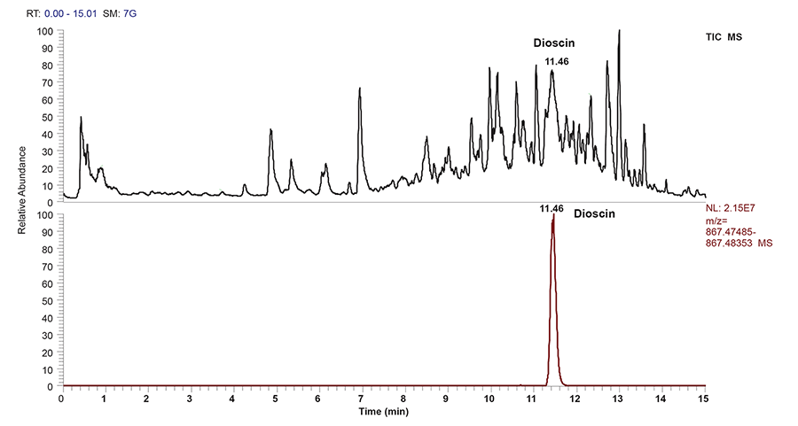
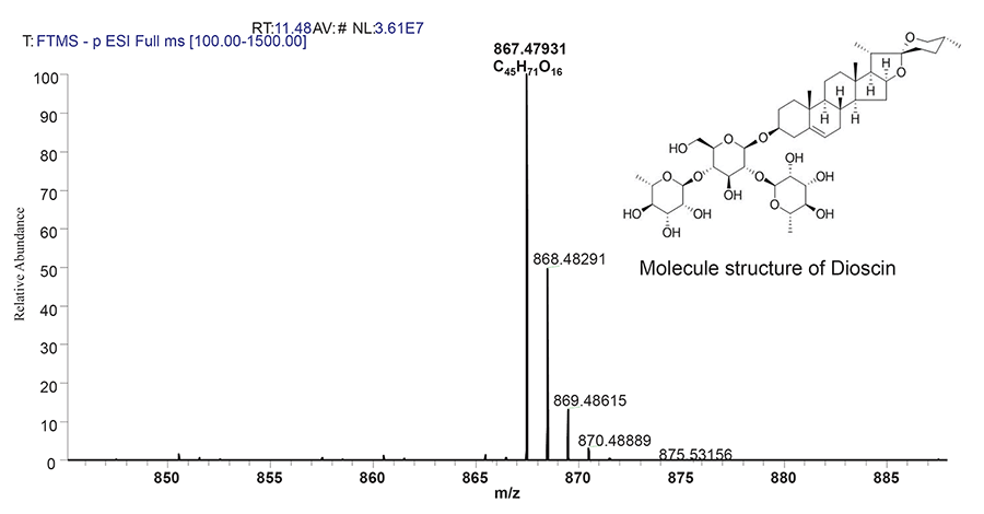
Ultrahigh performance liquid chromatography liquid chromatography (UHPLC) with MS detection has been used as an efficient tool for sample analysis and to facilitate structural elucidation. In the present study, mass specific detection was employed for identification of steroidal saponin due to its lack of specific chromophore for UV/VIS. Identification was performed using UHPLC-ESI-MS under developed chromatographic condition (Figure 1). Compound with a retention time of 11.48 min, produced a strong deprotonated molecule [M-H]- (m/z 867.47931) in (-)-ESI-MS. This compound is the main steroidal saponin present in the tuber and was tentatively identified as dioscin with structure molecule as shown in Figure 2 and Table 1. Rat Hepatotoxicity RT² Profiler™ PCR Array consists of the most relevant potential biomarkers for five major drug-induced hepatotoxic diseases such as cholestasis, steatosis, phospholipidosis, non-genotoxic hepatocarcinogenicity and necrosis, as well as generalized hepatotoxicity. Using real-time PCR, the expression of selected genes involved in hepatotoxic response with this array can be analyzed. Genes that consistently exhibit increased or decreased expression during these toxic responses in model systems serve as markers to predict potential adverse clinical outcomes.
No significant difference was observed at 250 and 1000 mg/kg b.w of D. hispida as compared to the control group (Table 2). However, several genes were significantly down and up-regulated upon treatment with D.hispida given at the highest dosage of 2000 mg/kg. Treatment of D.hispida at 2000 mg/kg b.w leads to significant upregulation of several genes such as Btg2 (32.22;0.02), Gsr (41.83;0.02), L2hgdn (20.52;0.05), S100a8 (51.28;0.03), Slc17a3 (2.24;0.02), Bhmt (1.85; 0.05), Cd68 (2.19; 0.02), Cyp1a2 (19.91;0.05) whereas several genes were downregulated such as Abcb1a (-29.31; 0.01), Aldoa (-3.45;0.05), Cdc14b (-4.93;0.03), Icam1 (-30.36; 0.02), Krt18 (-5.41; 0.03), Hpn (-4.33; 0.01) and Maob (-2.52; 0.04).
|
|
|
|
|
|
DISCUSSION |
|---|
Our study showed that the highest dosage of 2000 mg/kg D.hispida containing an active compound known as Dioscin caused mortality to the pregnant rats. The PCR Array indicated that several genes were found to be significantly upregulated and downregulated as the concentration of D.hispida given to the rats increased. This could possibly explain that the regulations of several gene expressions were affected by different concentrations of D.hispida given.
Genes such as Bhmt, Gsr, Slc17a3, Btg2 and Cyp1a2 were upregulated and based on the functional groups, these genes are classified in hepatoxicity group. Cd68 and L2hgdh are known to be involved in necrosis. S100a8 is categorized in phospholipidosis. In addition, several genes were significantly downregulated at the highest dosage and classified into different functional groups; Abcb1a and Icam1 are associated with cholestasis. Hpn is involved in phospholipidosis whereas Maob is associated with hepatoxicity. Aldoa and Krt18 are associated with nongenotoxic hepatocarcinogenicity. Cdc14b is involved in necrosis.
The presence of Dioscin in D.hispida may be associated with hepatoprotective effects as shown in previous studies. Dioscin has been shown to have a good protective effect against APAP-induced liver injury and this protective action is related to the regulation of mitochondrial function (17). In another study, Dioscin exhibited potent effects against liver fibrosis through the modulation of multiple targets and signaling pathways (18).
Plants containing active compounds such as glucosides, acids or alkaloids are used as medicines, however when taken in excess may lead to several adverse effects (19). A previous study indicated that administration of D. hispida given up to 1250 mg/kg crude extract in rats caused several poisoning symptoms, irregularities and complications to various organs including liver and kidney (15).However, in another study involving acute and sub-acute toxicity experiments, 50% of Benincasa hispida aqueous ethanolic extract given up to 2000 mg/kg to rodents did not cause mortality, behaviour changes and no significant alteration in hematological as well as biochemical parameters results indicating that there is a wide margin of safety for the therapeutic use (16).
The knowledge of gene expression patterns found in this study has demonstrated practical benefits in terms of predicting pathological events and toxic endpoints. Thus, gene expression data can provide an early indication of toxicity because toxin-mediated changes in gene expression are often detectable before clinical observation, clinical chemistry or histopathology (20–22).
|
|
CONCLUSIONS |
|---|
The consumption of D.hispida extract containing Dioscin at lower dosage below 2000 mg/kg may not be harmful. In response to administration of D.hispida extract given at the highest dosage, the alterations of several genes categorized in different hepatotoxic group functions were observed affecting the liver functions of pregnant rats. The hepatoxicity gene array can be used in developing mechanism-based biomarkers for diagnosis or prognosis of hepatotoxicity-related coagulation abnormalities in the early stage of drug development.
|
|
ACKNOWLEDGEMENT |
|---|
This study was supported by the National Institute of Health, Malaysia, under project number JPP-IMR 16-031. The authors gratefully acknowledge the Director General of Health, Malaysia and Director of the Institute for Medical Research, Kuala Lumpur, for permission to publish the finding of this study.
|
|
CONFLICT OF INTEREST |
|---|
The authors declare that there is no conflict of interest regarding the publication of this paper.
|
|
REFERENCES |
|---|