| REVIEW ARTICLE |
|
|
1 Irvine High School, 4321 Walnut Ave, Irvine, CA 92604, USA;
2 College of Veterinary Medicine, Western University of Health Sciences, Pomona, CA 91766, USA
Corresponding Author: Jijun Hao, College of Veterinary Medicine, Western University of Health Sciences, Pomona, CA 91766, USA. E-mail: jhao@westernu.edu.
Running title: STEM CELL-BASED SKIN REJUVENATION
|
|
ABSTRACT
|
|---|
Skin aging is a process in which the collagen and elastin in the dermis layer of human skin gradually get lost as time passes, resulting in wrinkles and lack of resilience of the skin. As people’s aesthetic standards have constantly grown in the recent years, many tried to eliminate wrinkles to improve their appearance; consequently, the demand for anti-aging has as well increased in the market. In the wide research for anti-aging, multipotent mesenchymal stem cells (MSCs) found in various sources including bone marrow, adipose tissue, umbilical cord and umbilical cord blood have been proved to eliminate wrinkles effectively by helping to promote collagen and elastin in the skin. In addition, MSCs also receive little or no response from human immune system, which is ideal for donor MSCs to be transplanted to other patients. In both preclinical and clinical studies, MSCs and their derivatives (such as MSC-derived conditioned medium and extracellular vesicles) have been proven to be beneficial in skin rejuvenation. This article specifically focuses on the recent advances in MSCs and MSCs-derivatives for skin rejuvenation.
KEY WORDS: Anti-aging; Skin Rejuvenation; Mesenchymal Stem Cells (MSCs); MSC-conditioned Medium; MSC-exosomes; Extracellular Vesicles|
|
INTRODUCTION |
|---|
The skin is the largest organ in the human body. It protects the body from external microbial agents, helps regulate body temperature, prevents physical injury, and enables touch sensitization (1). The skin consists of three layers, the epidermis (the top layer), the dermis (the middle layer) and the hypodermis (the bottom layer). The top epidermis acts as a barrier to protect the skin against bacteria, viruses, ultraviolet (UV) exposure and others (2). The middle layer dermis contains collagen and elastin, two critical proteins that makes skin cells strong, resilient, and flexible, and also helps the stretched skin regain its shape (3). In the process of aging, the production of collagen, elastin and other molecules in fibroblast cells decreases, resulting in a thinner layer of dermis (4). Meanwhile, due to an increase in the production of matrix metalloproteases (MMP) and oxidant activity in aged skin (4-7), collagen becomes disorganized, and elastin is degraded. Further, the process of skin aging increases the likelihood of diseases such as autoimmune disorders, dermatitis, eczema and melanoma (8).
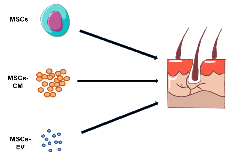
Since a significant number of people worldwide desire to slow down their skin aging and maintain youthful skin appearance, efforts have made to meet this requirement in the past decades, including developing topical drugs as well as invasive and non-invasive treatment procedures (5). In recent years, many studies have reported the applications of mesenchymal stem cells (MSCs) and their derivatives in improving the quality of the aging skin. MSCs are multipotent stem cells with ability to differentiate into several cell lineages including osteocytes, adipocytes and chondrocytes(9). MSCs were initially identified from bone marrow, and later from many other tissue sources including adipose tissue, umbilical cord and umbilical cord blood, etc (9). In addition, MSCs do not express major histocompatibility complex (MHC) Class II surface antigens and very low levels of MHC Class I antigens (10). Such unique MSCs immune tolerant features greatly facilitate transplanting the donor MSCs to a patient without inducing systemic severe immune rejection in the patient. Moreover, it has been widely accepted that MSCs actually exhibit their functions by the secreted growth factors or extracellular vesicles (EVs) in the conditioned media derived from MSCs. As a result, such cell-free MSCs-conditioned media (MSCs-CM) and EVs have been extensively studied for skin rejuvenation. In this article, I review the recent advancements in anti-aging skin rejuvenation by MSCs, MSCs-CM and MSCs-EVs (Figure 1).
|
|
|
PRECLINICAL STUDIES OF THE MSCS-BASED SKIN REJUVENATION |
|---|
Before the application of MSCs on human subjects for skin rejuvenation, multiple studies have been performed in preclinical animal models such as mice and rats. For instance, Kim and colleagues induced skin wrinkles in hairless mice by UVB irradiation for 8 weeks, and then injected human adipose-derived MSCs (AD-MSCs) subcutaneously into the mice (11). They found that the AD-MSCs at either 1 × 10⁴ and 1 × 10⁵ cells dramatically reduced skin wrinkles and increased dermal thickness and collagen contents (11). Besides UVB, D-galactose is also often used to induce aging mouse model (12). In 2014, Zhang examined the anti-aging effect of adipose MSCs in a D-galactose-induced aging mouse model by subcutaneous injections of 1x10⁶ adipose MSCs (13). They reported that adipose MSCs treatment reversed the aging phenotype and may contribute to the regeneration of skin during aging (13). In addition, bone marrow-derived MSCs (BM-MSCs) have examined for skin aging as well. In 2017, Liu et al reported that subcutaneous injection of BM-MSCs into rat subjected to D-galactose-induced aging markedly improved the D-galactose-induced histological abnormalities of the skin by ameliorating oxidative stress in aged skin (14).
|
|
PRECLINICAL STUDIES OF THE MSCS-CM-BASED SKIN REJUVENATION |
|---|
In the research by Kim and colleagues to study AD-MSCs for skin rejuvenation in mice (11), they also examined the impacts of the adipose MSCs-CM on dermal fibroblasts irradiated by UVB, and found that the adipose MSCs-CM decreased the UVB-induced fibroblast cell death and increased the collagen expression (11). Similarly, Kwon et al studied the conditioned medium derived from human bone marrow MSCs (BM-MSCs) in UVB-irradiated hairless mice (15). They demonstrated that the BM-MSCs-CM significantly increased pro-collagen synthesis and induced the repair of dermal damage and effacement of wrinkles on UVB-irradiated hairless mice (15). In 2014, Chen et al compared two types of the BM-MSCs-CM, hypoxic BM-MSC-CM (hypoCM) vs. normoxic BM-MSC-CM (norCM) in the skin wound recovery (16). They found that, in comparison to norCM, the hypoCM dramatically enhanced the cell growth of keratinocytes, fibroblasts and endothelial cells in vitro, accelerated skin wound recovery in mice (16). In addition to BM-MSCs-CM, adipose MSCs-CM was also studied for the protective activity in the UVB-induced skin aging in human keratinocytes and dermal fibroblasts (17). Li et al reported that adipose MSCs-CM could upregulate antioxidant response element and increase the expression of collagen synthesis enhancer gene in keratinocytes and dermal fibroblasts which are known to function as barriers to the “aging ray” UVB (17, 18). These studies highlight the potential of MSCs-CM for inhibiting UVB-induced wrinkle formation.
|
|
PRECLINICAL STUDIES OF THE MSCS-EVS-BASED SKIN REJUVENATION |
|---|
In recent years, interest in extracellular vehicles (EVs) derived from MSCs is emerging for their better safety and bioavailability. In 2020, Xu and colleagues studied effect of AD-MSCs-derived EVs (AD-MSCs-EVs) on mice with UV-induced photoaging (19). In this study, PBS (for control), 150 μg and 300 μg AD-MSCs-EVs were subcutaneously injected weekly into UVB-induced photoaging mice for 8 weeks to compare the effects of different amounts of EV injections on mice. The result showed that AD-MSCs-EVs dose dependently improved in the photoaging skin condition in mice by reducing intracellular reactive oxygen species production and promoting antioxidant enzyme expression (19). In 2021, a study of the EVs derived from human umbilical cord mesenchymal stem cells (hUC-MSCs-EVs) was reported, and the result showed that subcutaneous injection of hUC-MSC-EVs exhibited antioxidant and anti-inflammatory effects against UVB radiation-induced skin photodamage in rats (20).
Exosomes are a subtype of EVs with sizes ranging 30–150 nm in diameter (21). Kim et al investigated the functions of exosomes that are derived from hUC-MSC-CM in cutaneous collagen synthesis and permeation (22). They have shown that the hUC-MSC-Exosomes can reach the outermost layer of the epidermis after 3 hours and gradually approached the dermis after 18 hours and promote the synthesis of collagen and elastin after 3 days of treatment on human skin, supporting their potential for skin rejuvenation (22). In addition, Zhang et al conducted a study in in-vitro porcine skin and in-vivo mouse skin to determine whether marine sponge Haliclona sp. spicules (SHSs) could effectively improve the skin delivery of hUC-MSC-Exosomes (23). They found that SHSs dramatically increased in both porcine and mouse skin; the combination of the hUC-MSC-Exosomes with SHSs showed significant anti-photoaging effects in mice, including reduction of microwrinkles, alleviation of histopathological changes and improved expression of extracellular matrix constituents whereas hUC-MSC-Exosomes alone produced considerably weaker effects (23).
In summary, MSCs-EVs may replace MSCs and MSCs-CM as a potential agent for preventing or treating skin photodamage and skin rejuvenation.
|
|
CLINICAL STUDIES OF THE MSCS AND THEIR DERIVATIVES FOR SKIN REJUVENATION |
|---|
Given the promising outcomes from the preclinical studies, the clinical studies of MSCs-CM and MSCs-EVs have been initiated recently.
In 2018, Kim et al investigated a cream of the hUC-MSC-CM on dermal density and wrinkles in 22 human patients (24) (Table 1). After daily use for four weeks, it was found that the cream improved skin density by 2.46% and reduced the skin wrinkles of eye-end area dramatically (24). Recently Liang and colleagues conducted the combination of hUC-MSC-CM and micro-needling in skin rejuvenation in 28 volunteers with facial skin aging (25) (Table 1). Their left and right sides of the faces were randomly applied with saline or the hUC-MSCs-CM via micro-needling for five sessions at two-week intervals. The condition of skin brightness and texture were then evaluated by dermatologists, and a self-evaluation questionnaire were recorded as well. The result of the study showed that hUC-MSCs-CM-plus- micro-needling treatment statistically improved skin brightness and skin texture significantly in contrast to micro-needling saline alone (25). In addition, no severe side effects were reported during the whole study period (25). Similarly, El-Domyati et al compared the effect of amniotic fluid MSCs derived conditioned media (AF-MSC-CM) combined with skin needling versus the needling alone in the treatment of facial aging (26) (Table 1). Their recruited 10 volunteers suffering from skin aging and treated them with skin needling on both sides of faces two weeks apart for five sessions. After skin microneedling, AF-MSC-CM was applied topically to the right side only, and the clinical outcomes were then assessed at 1 month after the last session. Their result indicated that the right side of the face, which was treated with both microneedling and AF-MSC-CM, was significantly improved regarding skin aging comparing to the left side, which was treated with microneedling alone (26).
In consistence, Bhat et al conducted a trial by applying a topical formulation containing human BM-MSCs-CM to 20 Indian female subjects twice daily for the duration of 12 weeks (27) (Table 1). The efficacy was evaluated at 4 weeks and 12 weeks after the treatment by examining clinical parameters such as eye puffiness, radiance, skin smoothness, even skin tone, periorbital fine lines and wrinkles, crow’s feet, whitening, pigmentation, skin tightening, and refreshing/soothing effect. The result demonstrated that the majority of the clinical parameters were improved after 4-week treatment and continued to improve at 12 weeks after the treatment (27).
In addition, exosomes isolated from AD-MSCs-CM were examined in human subjects for skin rejuvenation as well. Cho et al assessed the skin brightening efficacy of a cosmetic formulation containing AD-MSCs-exosomes in 21 female volunteers with hyperpigmentation in a split-face, double-blind, randomized placebo-controlled study. The formulation of the AD-MSCs-exosomes statistically decreased the melanin contents compared to the placebo control and a further improvement was observed for the transdermal delivery of the AD-MSCs-exosomes (28) (Table 1).
In summary, both MSCs-CM and MSCs-exosomes exhibits anti-aging efficacy, and they could be used for facial rejuvenation.
|
|
|
CONCLUSION |
|---|
Skin aging is a natural process involving the decrease of skin collagen and elastin synthesis which lead to the disorganization of connective tissue. Currently skin rejuvenation is in a very high demand. MSCs are multipotent stem cells which can be isolated from various sources including bone marrow, adipose tissue, umbilical cord and umbilical cord blood. Given their unique immune tolerant features, cumulative studies have shown that MSCs treatments have great beneficial effects for skin rejuvenation. Subsequently, it was proven that MSCs exhibit their functions by the secreted growth factors in the MSCs-CM. As a result, the cell-free MSCs-CM and the CM-derived EVs have been demonstrated to greatly slow down skin aging and promote skin rejuvenation. However, before MSCs and their derivatives can be widely used in clinical practice for skin rejuvenation, some challenges need to be overcome. For example, standardized protocols are still lacking for isolation, culture and expansion of MSCs from various sources. In addition, delivery dosage and administration route are thought to affect the heterogeneity of MSCs as well, and therefore, more and larger randomized blind controlled trials are needed to optimize MSC treatment protocols. In recent years, the EVs and exosomes isolated from MSCs are believed to be superior over MSCs in skin rejuvenation as they have several advantages including higher safety profile, lower immunogenicity and the ability to easily cross biological barriers. However, the use of MSC-EV and their exosomes in clinical settings is facing some challenges such as no effective larger scale production methods available and lack of quantification and characterization (29). Despite those issues, MSCs and their derivatives holds a great promise in skin rejuvenation.
|
|
CONFLICT OF INTEREST |
|---|
The author discloses no conflict of interest.
|
|
REFERENCES |
|---|