| Original Article |
|
|
1Department of Pharmaceutical Biochemistry, Laboratory of Molecular Biology and Pharmacogenomics, Łódź, Poland;
22Department of Surgery District Hospital, Łęczyca, Poland 18, 07743 Jena;
Corresponding author:Marek Mirowski, Department of Pharmaceutical Biochemistry, Laboratory of Molecular Biology and Pharmacogenomics, Muszynskiego 1 Street, 90-151 Lodz, Poland. Tel: +48 42 677-91-30; E-mail: mirowski@ich.pharm.am.lodz.pl.
| |
ABSTRACT |
|
|
INTRODUCTION |
|
|
MATERIAL AND METHODS |
|
|
RESULTS |
|
|
DISCUSSION |
|
|
ACKNOWLEDGEMENTS |
|
|
REFERENCES |
|
|
ABSTRACT
|
|---|
P65 gene expression level was determined in colon cancer cases by means of real-time PCR. 51 cases of colorectal carcinomas showing positive RT-PCR signals for P65 gene expression selected from 109 frozen samples were further investigated by quantitative real-time PCR. P65 levels were higher in cancer with metastases to lymph nodes and distant metastases. Higher levels were observed in more advanced cases classified as III and IV according to pTNM classification. In two groups of patients with vessel invasion and absence of lymphocytes in tumour tissue, the presence of P65 expression correlated with shorter survival time. Quantitative results confirmed that P65 gene expression in colon cancer is engaged in the process of metastasis formation and could be correlated with worse prognosis for the patients.
KEY WORDS:
colon cancer; P65 gene; real-time PCR
|
|
INTRODUCTION |
|---|
A 65 kDa protein (P65) is expressed by many types of tumour cells. P65 was originally isolated from the MCF-7 human breast cancer cell line (1). The presence of the 65 kDa protein was observed not only in tumour tissue but also in the sera of patients diagnosed with different types of neoplasm including colon cancers (2-6). Blood levels of P65 in patients suffering from cancer were significantly higher in comparison to healthy men. It was also showed that in colon cancer, estimation of P65 level in serum parallel to CA19-9 and CEA improved the threshold of cancer detection when compared to CEA or to CA19-9 levels alone (7). Such results were obtained with the use of anti-P65 monoclonal and polyclonal antibodies raised against this protein (5, 6, 8). Immunohistochemical analysis performed in cases of breast carcinomas revealed both cytoplasmic and nuclear localization of this protein in the tumour cells (9).
Qualitative analyses of P65 gene by reverse-transcriptase PCR were performed in various types of leukemia (2), breast (10) and prostate cancers (3). In breast cancer P65 expression was generally connected with small tumors without metastases in regional lymph nodes, while the absence Absence of P65 expression was observed in cases classified as fibroadenoma (10). Similarly, in the prostate cancers P65 was mainly present in well-differentiated tumors. These results suggested that expression of P65 may correlate with a better prognosis for these patients (3, 10). In contrast, in a study of a population of 109 colon cancer cases the results were quite different. In this group P65 gene expression correlated with grading, clinical staging and other histological features (4). Unexpectedly, significant statistical correlations between the presence of P65 gene expression and the depth of tumor invasion (T3, T4), presence of lymph node metastases (N1-N2) and distant metastases (M1) with high clinical stages (C2, C3 and D according to Astler-Coller classification) and with vessel invasion were found suggesting a poor prognosis (4). In this paper, gene expression was analyzed quantitatively by means of real-time PCR for the 51 colon cancer samples that had previously tested positive for P65 expression.
|
|
MATERIAL AND METHODS
|
|---|
Human colon cancer tissues
Tissue specimens of colorectal carcinomas were obtained from the Oncological Centre of Lodz, Poland under the license of the local ethics committee (KE/286/05). 51 cases for quantity analyses were chosen from the whole population (n=109) on the basis of analysis carried out by quality multiplex reverse transcriptase PCR (4). In this group, 23 cases of carcinomas were from women (mean age 60.5 ± 5) and 28 cases were from men (mean age 62.5 ± 5).
Quantification of the level of P65 gene expression
by real-time–PCR
For quantitative analysis, RNA was isolated using a Total RNA Prep Plus Minicolumn Kit (A&A Biotechnology, Poland) based on RNA isolation methodology developed previously (11). All RNA samples were treated with DNAse (Sigma) to remove genomic DNA. RNA quantity was calculated after measurement of absorbance. Equal amounts of RNA for each sample were transcribed into cDNA using Enhanced Avian HS RT-PCR Kit (Sigma). Real-time PCR was performed using iCycler (Bio-Rad) and SYBR Green JumpTM Start Tag ReadyMixTM (Sigma) according to manufacturer’s instructions.
The P65 primer set 5’-GGTCCACGGCGGACCGGT-3’ (forward) and 5’-GACCCCGAGAACGTGGTGCGC-3’ (reverse) and conditions used in the assay were previously described (10). The following reagents were used for thermocycling: 25 ml Jump Start Tag Ready Mix, 0.5 ml reference dye, 1 ml of forward primer (final concentration 0.2 mM), 1 ml of reverse primer, 2 ml magnesium chloride (final concentration 25 mM), 5 ml template cDNA, and water (final volume 50 ml.) Cycling parameters were hot starting at 98°C for 5 min, followed by 2 min initial denaturation at 94°C, followed by 35 cycles consisting of denaturation at 94°C, 1 min annealing at 54°C and 3 min extension at 72°C, followed by 7 min final extension at 72°C. As a loading standard, the expression of β-actin gene was quantified for each sample using 5’-GTGGGGCGCCCCAGGCACCA-3’ (forward); 5’-CTCCTTAATGTCACGCACGATTTC-3’ (reverse) primer set (12). The experiments with P65 and β-actin were done not as a multiplex, but in the separated tubes during the same PCR. Because of this the final results are not given as a relative ratios. The level of P65 was monitored by measuring the increased fluorescence of SYBR Green through the PCR cycles and estimated at the threshold cycle for each analysis. P65 expression levels were calculated on the basis of a standard curve (units used for preparing curve ng/ml) that was obtained by plotting known quantities of genomic P65 DNA. Then data were transformed into Microsoft Excel. Experiments for all samples were carried out in triplicate.
Statistical analyses were performed using the U Mann-Whitney test. Time-to-event distribution for survival in the whole population of 109 patients was estimated using the Kaplan-Meier method. The F-Cox test was used to test for differences in time-to-event distribution.
|
|
RESULTS
|
|---|
P65 gene expression levels
are related to clinico-histological
features
109 of colon cancer cases were analyzed by conventional reverse transcriptase PCR, 51 of which the presence of P65 gene expression. All positive cases were taken for further quantitative analysis using real-time PCR.
P65 gene expression levels were compared with several clinicopathological parameters such as depth of tumor invasion (T), lymph node metastases (N), and distant metastases (M) (TNM classification).
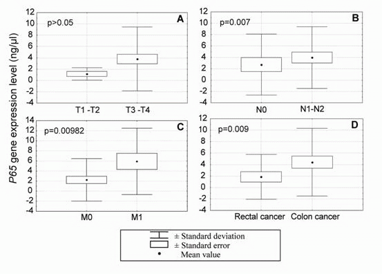
P65 gene expression in the more advanced tumors (T3 and T4, deep wall penetration) was 4 ng/ml, while in the T1-T2 group lower levels of expression were recorded (1 ng/ml). No statistically significant correlation was observed between P65 gene expression and tumor invasion depths (Figure 1A).
P65 gene expression was analyzed in cases with and without lymph node metastases. In cases without lymph node invasion (N0) P65 levels were 3 ng/ml, while in cases with lymph node metastases (N1-N2) P65 mRNA levels were 4 ng/ml (Figure 1B). This difference was statistically significant (p=0.0395 U Mann-Whitney test).
In carcinomas with distant metastases the levels of P65 expression were two times higher (M1: 6 ng/ml) than in the group of cancers without distant metastases (M0: 3 ng/ml) -Figure 1C. These differences were statistically significant (p=0.009872 U Mann-Whitney test).
P65 levels were statistically lower in cancers originating in the rectum as compared to those originating in the colon (p=0.009 U Mann-Whitney test, Figure 1D).
Comparison of P65 gene expression levels with pTNM staging showed that the more advanced cases (stages III and IV) had statistically significant higher levels of P65 (4 ng/ml) than stages I and II (2 ng/ml) - p=0.0218 U Mann-Whitney test.
The cases without lymphocytes in tumor tissue showed higher levels of P65 (4 ng/ml) then those with the presence of lymphocytes (2.5 ng/ml), but this difference was not statistically significant. Furthermore, in the group of tumors without vessel invasion the level of P65 gene expression was lower (2 ng/ml) than in tumors with the presence of vassel invasion (4 ng/ml) but this difference was also not statistically significant. The data describing P65 gene expression in relation to clinico-histological features are summarized in Table 1.
P65
expression
and survival time
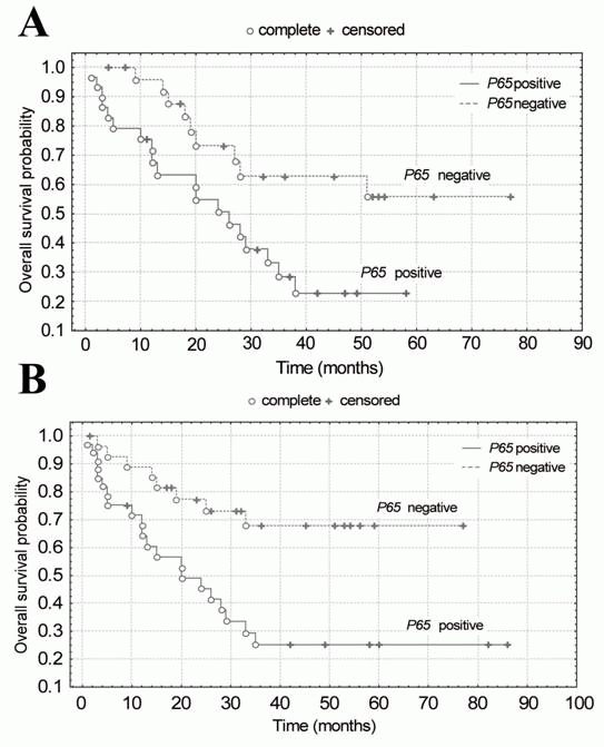
In the entire investigated population (n=109 patients), there was no statistically significant different in survival time comparing patients with and without P65 gene expression in spite of the visible tendency for longer survival of those without P65 gene expression (p>0.05). It is interesting to note that in the group of patients with vessel invasion (p=0.01396 F-Cox test, p=0.00267 log. rang test) and also in the group with the absence of lymphocytes in tumour tissue (p=0.02380 F-Cox test, p=0.01147 log. rang test) the presence of P65 expression was correlated with shorter survival time (Figure 2A and B). Further analysis of different variants didn’t show differences in survival time between P65 positive and negative groups.
P65 gene expression level in cancer and adjacent, potentially healthy
tissues
It may be of interest that 19 out of 51 investigated cases revealed P65 expression in quality analysis in both neoplastic and adjacent, potentially healthy colorectal tissue. Quantitative studies showed that in adjacent, healthy tissue P65 levels were statistically lower in comparison to cancer tissue (p=0.019).
|
|
|
DISCUSSION
|
|---|
The
potential role of P65 protein in cancer diagnosis and prognosis
Cancer of the large bowel is a serious and growing medical problem. Thus, there is a real need to identify factors engaged in development of colorectal cancer that could be useful in prognosis and therapy selection. The P65 gene and its tumour-associated P65 protein seem to be involved in neoplastic transformation and tumour progression also in colon cancer (4, 7). The pathways involved are still unknown and the biological functions of P65/P65 are not completely known and understood. On the basis of established partial amino acid and nucleotide sequences, homology to steroid receptors was revealed, especially strong in their DNA-binding domains (1, 13). However, other regions of the sequence do not show similarities. This suggests that P65 may be either a new receptor with unknown ligand or a transcriptional factor. Interaction of P65 with DNA can modulate transcription of some genes important for regulation of growth, development or cell differentiation (14, 15). This hypothesis is supported by the cytoplasmic and nuclear staining by anti-P65 antibodies (9). Immunohistochemical analysis showed that P65 cytoplasmic reactivity correlated with high levels of high estrogen and progesterone receptors level and with low clinical stage and grade. Nuclear manifestation of P65 expression was connected with more advanced stages, particularly with node and distant metastasis and high grade (9).
The potential role of P65 gene expression in
cancer diagnosis and prognosis
Qualitative studies using reverse transcriptase PCR showed that P65 gene expression is not present in healthy tissues but can be found only in cancer tissues or in benign changes that can progress to malignant tumours (3, 10) These data indicated that expression of this gene is connected with neoplastic transformation, but not in all investigated cases, e.g. colon cancer where nearly half of carcinomas expressed P65 (4). In these cases, the presence of P65 was connected with metastases to lymph nodes and distant metastases, and these correlations were statistically important. Such results were confirmed in the current study by real-time PCR analysis, where the levels of P65 were higher in more advanced cancers. It may prove P65 gene may play important roles in metastatic progression of colorectal cancer that could be potentially useful in clinical practice. The most important is the correlation between high levels of P65 and distant metastases. It should be noted that the cause of death in the colon cancer patients is often not the primary tumour but the secondary metastases to the liver. These changes are present in a clinically non-detectable form, known as micro-metastasis, in 50% of colorectal cancers cases at the time of diagnosis. For the patients with high levels of P65 without clinically manifested metastases more aggressive therapy may be indicated, and they should be monitored more closely.
P65 gene
expression level as an early indicator of cancer formation
It should be noted that in 19 out of the 51 cases investigated P65 was detected in both cancerous tissue and adjacent, healthy mucosa (classified by pathologist). Quantitative results showed significantly higher levels of P65 in cancer tissue. Nearly 80% of these 19 cases were classified according to pTNM as III or IV. Such results may suggest that P65 appeared earlier then typical neoplasmic changes visible to the histopathologist and that the margin of surgical intervention should be increased. This is in agreement with previous experimental studies where P65 rapidly accumulated in the blood plasma of rats 14 days serum after partial hepatectomy and treatment with N-diethylnitrosamine (DEN) and reached a plateau by 3 weeks. These changes were earlier than any neoplastic minifoci in hepatoma visible by histopathologists (16).
Although, it is too early to speculate about the molecular mechanisms of P65/P65 action and its role in cancer formation and spreading, this study shows that high levels of P65 gene expression correlate with advanced stage of disease in colon cancer. These results are in agreement with our earlier observation in breast cancer patients (9) where the presence of P65 in cancer cell nuclei also correlated with formation of metastases. Certainly, P65 protein and P65 gene expression may behave in different ways in various types of tumors but their presence and elevated levels may have prognostic significance. However, further investigations are needed to establish the P65 gene structure, its genome localization, its role in tumor development and any diagnostic/predictive value of P65/P65 in different cancers.
|
|
|
|
ACKNOWLEDGEMENTS
|
|---|
Special thanks to BioRad representatives that gave us opportunity to use iCycler Real-Time PCR detection system.
|
|
REFERENCES
|
|---|