| ORIGINAL ARTICLE |
|
|
1 Department of Pediatric, Faculty of Medicine, Cairo University, Cairo, Egypt;
2 Department of Pediatric, National Research Centre, Cairo, Egypt
3 Department of Clinical & Chemical Pathology, National Research Centre, Cairo, Egypt
Corresponding Author: Manal F. Elshamaa, Pediatrics Department & Medical Research Centre of excellence, National Research Centre, Cairo, Egypt; Elbohous street, Dokki, cairo Egypt. Postal code 12311 E-mail: manal_elshmaa@hotmail.com.
| |
ABSTRACT |
| INTRODUCTION | |
|
|
METHODS |
|
|
RESULTS |
|
|
DISCUSSION |
|
|
CONCLUSION |
|
|
CONFLICT OF INTEREST |
|
|
ACKNOWLEDGMENTS |
|
|
REFERENCES |
|
|
ABSTRACT
|
|---|
Background: The equilibrium between regulatory cells and cytotoxic cells may define graft consequence. We investigated the relationship between the expression of main regulatory and cytotoxic markers (i.e., FOXP3 and granzyme B (GZM-B), respectively) and acute rejection (AR) in the peripheral blood of pediatric renal transplant recipients.
Methods: In this retrospective study, FOXP3 mRNA expression and serum GZM-B levels in peripheral blood samples from 47 first-time pediatric kidney-transplant recipients were measured, with 17 children classified as possessing AR; whereas the remaining 30 children had functionally stabilized allografts.
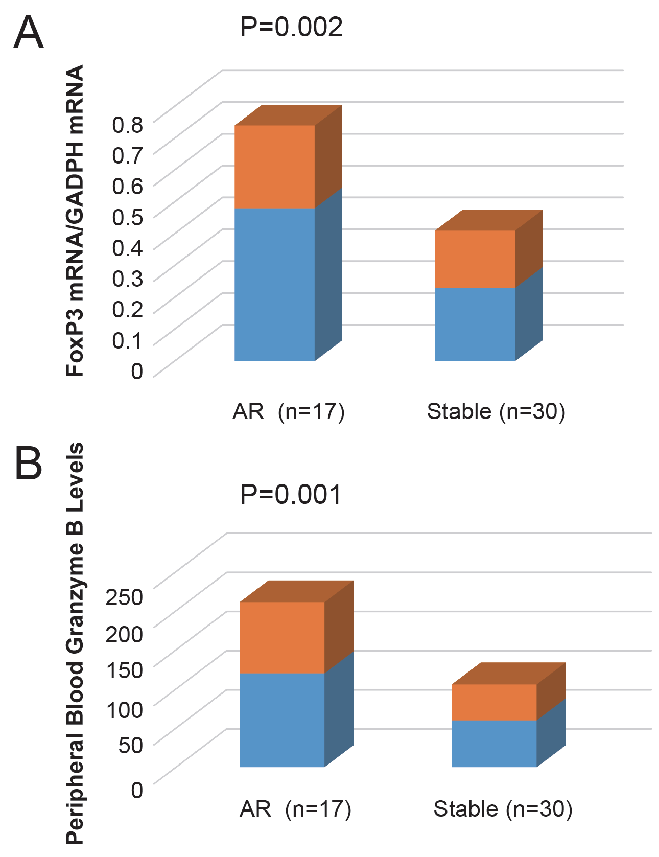
Results: Levels of the FOXP3 mRNA vs. the expression levels GADPH mRNA (FOXP3 mRNA/GADPH mRNA) were significantly elevated in children with AR than those with stabilized renal allograft (0.48 ± 0.26 vs.0.23 ± 0.18, respectively, P=0.002) Also, serum GZM-B levels in the AR group were elevated than those in the functionally stabilized children (120.07 ± 91.42p g/ml and, 60.16 ± 46.29 pg/ml respectively, P=0.01). ROC curve evidenced that measuring FOXP3 mRNA may have a scope as a decision-taking agent in clinical proceedings to diagnose AR. Measuring peripheral blood FOXP3 mRNA elucidated scope to help in the noninvasive diagnosis of AR.
Conclusions: Our results emphasize FOXP3 mRNA as a biomarker for AR in pediatrics. Assessment of regulatory/cytotoxic profiles in the peripheral blood of pediatric renal transplant recipients is a potentially useful tool for patient selection and early detection of rejection. Depending on many variables, such as the method of sample normalization, the technique used, the extent of graft inflammation, the immunosuppression regimen, depletion/ repletion of T-lymphocyte component, the importance of FOXP3 may differ.
|
|
INTRODUCTION |
|---|
End Stage Renal Disease (ESRD) has a mischievous effect on the patient's life better and though dialysis is one of the most forms of renal replacement therapy yet it has deleterious consequences; exposure of the child to infections, provocation of cardiovascular complications and bone diseases as well as growth retardation. The advances in the field of dialysis do not improve the increasing mortality rate among ESRD cases (1). Renal transplantation offers a better quality of life and prognosis for these patients. It has been reported that 3 months and 1 year remaining alive of transplanted kidneys from living donors are 99.5% and 98.5%, respectively (2).
Graft with perfect function and immunosuppressive doses reduction to the maintenance dose with the minimal complications are the targets of transplantation medicine. Therefore, knowing the optimum immunological and cellular mechanisms of the allograft rejection process and development of strategies for stimulating tolerance and decreasing the specified immune responses due to transplant antigens are critical in this respect (3).
Rejection of transplanted tissues occurs as a consequence of the interplay between mechanisms that keep up the tolerance to the graft and factors that promote rejection. One of the most significant causes of graft loss is acute rejection (AR), involving both cellular and/or humoral immune responses 4. Cellular rejection is morphologically characterized by cytotoxic T cells (4, 5) infiltrating the graft and promote apoptosis of renal tubular epithelial cells through two mechanisms; the first is exocytosis of cytotoxic granules containing granzymes A and B delivered into a target cell via transmembrane pores formed by perforin. The second is death receptor-induced apoptosis e.g. CD95 ligand/CD95 pathway. Humoral rejection is associated with complement cascade activation, vasculitis and immunoglobulins deposition (6). T lymphocytes implicated not only in the initiation of the cascade of mechanisms implying rejection but also share in mechanisms that keep up graft tolerance (7).
Achievement of transplantation clinical tolerance has been a noteworthy objective with intensive research in this field for more than 50 yrs. (8, 9). In rodents, allograft tolerance is repeatedly linked with regulatory T cells (Tregs) (distinctive population of suppressor T cells) (10). The constitutive expression of the forkhead-winged helix transcription factorFOXP3 on CD4+CD25++ Tregs causes them to be differentiated from other cell types (11). A transcriptional repressor FOXP3 is essential not only for Tregs development but also its function and could be utilized as a biomarker for Tregs (12). Antigen-stimulated T-cell proliferation is suppressed by these inherently active CD4+CD25+FOXP3+ Tregs by suppressing cytokines of T helper (Th)-1 released from T-cell population effector. Repression utmost probably happened by wherewithal of a mechanism -depends on cell-to-cell communication, likewise by the Tregs anergic phenotype in vitro and their disability to secrete interleukin-2 (IL-2) (13, 14). Human studies affirmed that FOXP3 cells seem to be an inherited ingredient of AR, with elevated FOXP3 both in the urine (15) and the graft (16). Thus, the outcome of the autograft possibly depends on the balance between Teff (effector cells) and Tregs ratio, wither quantitative or qualitative predominance. Acute allograft rejection is also associated with granzyme B (GZM-B) expression by mononuclear cells in the graft and GZM-B was declared as a trustworthy peripheral blood, urine, and intragraft biomarker of AR (17, 18).
Aim: we aimed to investigate the balance between regulatory cells (FOXP3) and cytotoxic cells (GZM-B) in acute rejection in pediatrics kidney-transplant recipients.
|
|
METHODS |
|---|
Subjects and Study Design
Design. All the patients’ guardians gave informed written consents before participating in the study, which was approved by the Ethics Committee of NRC and PNU in Egypt.
Setting and population. Forty-seven children with ESRD who had received an allograft at Pediatric Nephrology Unit (PNU), Abu el Rish Children’s Hospital, Cairo University, Egypt, were evaluated in this study as well as 20 healthy age-matched, unrelated controls (12 males, mean age 8.7 ± 4.51 yr). Healthy children were recruited from the Pediatrics Clinic of the Medical Research Centre of Excellence (MREC), National Research Centre (NRC), Egypt. The study began April 2013 and ended December 2013. The time elapsed from the time of transplantation to the point of the study was 2.39 ± 0.97 yrs (range 0.5-4.5 yrs).
All patients had received their first transplantation and followed at regular intervals at the transplantation clinic. Baseline demographic characteristics, age, gender, etiology of end-stage renal disease (ESRD), treatment modality and duration before transplantation, details of the transplantation procedure, immunosuppressive regimens, as well as the time between specimen collection and kidney transplantation were recorded.Setting and population. Forty-seven children with ESRD who had received an allograft at Pediatric Nephrology Unit (PNU), Abu el Rish Children’s Hospital, Cairo University, Egypt, were evaluated in this study as well as 20 healthy age-matched, unrelated controls (12 males, mean age 8.7 ± 4.51 yr). Healthy children were recruited from the Pediatrics Clinic of the Medical Research Centre of Excellence (MREC), National Research Centre (NRC), Egypt. The study began April 2013 and ended December 2013. The time elapsed from the time of transplantation to the point of the study was 2.39 ± 0.97 yrs (range 0.5-4.5 yrs).
All patients had received their first transplantation and followed at regular intervals at the transplantation clinic. Baseline demographic characteristics, age, gender, etiology of end-stage renal disease (ESRD), treatment modality and duration before transplantation, details of the transplantation procedure, immunosuppressive regimens, as well as the time between specimen collection and kidney transplantation were recorded.
Creatinine was measured at least monthly post-transplant. The patients were divided into two groups based on the presence or absence of acute rejection: 17 patients suffered from acute rejection and 30 had stable allograft function. Peripheral blood samples were collected after kidney transplantation, at the time of rejection.
Diagnostic criteria for acute organ rejection were: sudden decrease in urine output, fever, and abdominal tenderness accompanied by increased serum creatinine and urea nitrogen decreased or unchanged urine specific gravity, hematuria and proteinuria. In addition, ultrasound examination showing increased kidney volume (with or without decreased blood flow), and an increased blood flow index. Acute rejection which is cellular rejection due to T cell activation encountered in the first week after post-transplant was defined and graded according to the Banff Criteria (19). It was defined as either borderline/suspicious or acute rejection in patients with stable serum creatinine values at the time of biopsy [grades 3 and 4] (20). No protocol biopsies were performed, particularly as the patients were pediatric patients where invasive biopsy accrues more cost and risk than in the adult population. Renal biopsies were only performed if there were clinical indications with suspicion for allograft dysfunction.
Organ recipients showing signs of chronic calcineurin inhibitor (CNI) nephrotoxicity, acute tubular necrosis, ureteral obstruction and/or renal artery stenosis of the graft, arterial and venous thrombosis, and infection-induced fever were excluded from the study. 17 patients were diagnosed with organ rejection and the diagnosis confirmed by renal graft biopsies. The timing of acute rejection in our patients ranged from one day to 16 months post-transplantation, with 52% of rejections occurring in the first 6 months post-transplantation.
The initial FK506 dose was 0.16 mg/kg per day by oral route (1.5-6 mg/day), and target trough levels were 3-14 ng/ml in the first 3 months and 4.5 ng/ml in the FK506/everolimus group. The initial dose of mycophenolate mofetil (MMF) was 360-1440 mg/day, and the dose was modified based on adverse effects such as diarrhea or leucopenia. IL-2 receptor blocking antibody (anti-IL-2R Ab, Basiliximab) (Simulect, Novartis Pharmaceuticals, Basel, Switzerland), was given to 10 patients (BSX group) (CsA or FK506 based immunosuppression) 4 hrs before and 3 days after renal transplantation (two 10 mg doses for patients weighing less than 35 kg, and two 20-mg doses for patients weighing more than 35 kg). Anti-thymocyte globulin (ATG) (Thymoglobulin_, Genzyme Transplant, Cambridge, MA) was given to 29 patients (THYMO group) as a single dose of 5-8 mg/kg on transplantation day (Day 0). Everolimus was administered 2 mg per day and Sirolimus has loaded 6 mg per day and then adjusted the dose of 2 mg/day was maintained with target trough level of 5-15 ng/ml.
Data collection
In kidney transplantation, the blood sample was withdrawn after transplantation. In patients having episodes of acute rejection, the samples were taken during the period of rejection and the genotypes and the assay were performed.
Soluble Granzyme B (GZM-B) assay
Serum levels of GZM-B semi-quantitative measurement were done by in vitro ELISA Kit (Ray Biotech, Inc., USA) in the laboratories of Clinical and Chemical Pathology Unit, of NRC in Egypt.
Detection of FoxP3 mRNA using real-time quantitative PCR
Total RNA was extracted from 2 ml anticoagulated whole blood using GeneJET whole blood RNA Purification Mini Kit (Thermoscientific#K0761).The quality of the isolated RNA was evaluated spectrophotometrically. The purified RNA has an A260/280 ratio between 1.9-2.1. First strand cDNA was synthesized according to the standard protocol of Revert Aid First strand cDNA synthesis kit (Thermo-scientific #K1621, #K1622). FOXP3 mRNA levels were quantified by real-time PCR with the Thermo-scientific PIKO24 Real-Time PCR detection system-THERMOFISHER Scientific Inc. MA, USA. FOXP3 specific primers and a fluorescent TaqMan probe were designed as follows; FOXP3 Primers: FOXP3 Primers: 5'- CAGCACATTCCCAGAGTTCCTC-3’ and 5’-GCGTGTGAACCAGTGGTAGATC-3' .FOXP3 Probe: 5'-FAM- TCCAGAGAAGCAGCGGACACTCAATG-TAMRA-3’. Pre-developed TaqMan Assay Reagent for human GAPDH (20x pre-mixture of GAPDH specific primers and an internal fluorescent TaqMan probe) was utilized for measurement of GAPDH mRNA levels as an internal control. Each PCR sample contained 0.6 um primers and 0.2 um TaqMan probe in a final volume of 25 UL and amplification was carried out via 15 min. at 95 C denaturation step followed by 45 cycles of 15 sec. at 94 C and 60 sec. at 60 C.
Data Analysis
Statistical analyzes were performed by SPSS 16.0 computer program and Pearson’s Chi-Square test. Data were summarized as mean ± SD, range or percentage. Histograms and normality plots were used for evaluating the normality of data. For those data with the skewed distribution, log transformation was performed before a t-test. Data were evaluated between the experimental groups by independent t-test. The diagnostic accuracy of the biomarkers for diagnosis of infection was expressed as the area under the corresponding receiver operating characteristic curve (AUROC) and the respective areas under the curves were calculated with 95 % confidence intervals. Calculated statistical power was 0.942(94.2%) using pass version 11. Multiple regression analysis was performed to assess the influence of the regulatory/cytotoxic T cell profiles on AR. A p value of <0.05 was considered statistically significant.
Procedure. We investigated the balance between regulatory cells and cytotoxic cells in acute rejection in pediatrics kidney-transplant recipients.
|
|
RESULTS |
|---|
Comparisons of the clinical and biochemical characteristics of the studied groups
The two patient groups did not significantly differ as regards to age, gender, the HLA mismatch rate, or cold ischemia time. Children in both groups received MMF (2-1500 mg/m2) and corticosteroids (prednisone 2-8 mg/d), the doses used did not significantly differ between in each group. The cyclosporine A/FK506 ratio was 16/12 in the performance stabilized allograft recipient and 2/12 in the AR children; the plasma trough concentrations did not significantly differ between the two groups (Table 1).
Peripheral blood levels of FoxP3 mRNA expression of allograft recipients in the studied groups
Post-transplantation, FoxP3 mRNA levels peripheral blood in vs. GADPH expression levels (FOXP3 mRNA/GADPH mRNA) in the AR group were highly significantly elevated than those in performance the stabilized children (0.48 ± 0.26 and 0.23 ± 0.18, respectively, P=0.002) (Figure 1A).
Comparisons of granzyme B levels in the peripheral blood of children of allograft recipients in the studied groups
Post-transplantation, peripheral blood granzyme B levels in the AR group were significantly higher than those in the functionally stabilized children (120.07 ± 91.42 (pg/ml) and, 60.16 ± 46.29 (pg/ml) respectively, P=0.01) (Figure 1B).
To distinguish AR from infections, we compare children with positive cytomegalovirus (CMV) infection with those with a negative one, we found that infectious complications such as CMV infection also did not lead to any significant changes in either of regulatory FoxP3 mRNA expression or cytotoxic GrzB levels. (FOXP3,0.32 ± 0.25vs.0.30 ± 0.20 (FOXP3 mRNA/GADPH mRNA), p=0.86. GrzB, 78.64 ± 70.05pg/mlvs.55.05 ± 35 pg/ml.14, p=0.43).
The influence of immunosuppressants on the regulatory (FOXP3) and cytotoxic (GrzB) profiles in the peripheral blood pediatric kidney-transplant recipients
When analyzing the various study groups, the FOXP3 mRNA/GADPH mRNA expression between the patients' groups was not statistically different; they were found to represent 0.38 ± .25 in FK group (n=24) and 0.27 ± 0.23 in CsA group (n=18) (P=0.21). Also, serum GZM-B levels between patients groups were not statistically different; they were found to represent 82.95 ± 79.76 pg/ml FK group and 73.14 ± 50.98 pg/ml in CsA group (P=0.86).
The influence of induction immunosuppressants on the regulatory (FOXP3) and cytotoxic (GZM-B) profiles in the peripheral blood pediatric kidney-transplant recipients
Thirty-nine patients received induction therapy following one of two protocols: 1) THYMO group (n =29) and BSX group (n=10). Comparing the FOXP3 mRNA/GADPH mRNA expression between patients groups was not significantly different; they were found to be 0.28 ± 0.21 in THYMO group and 0.48 ± 0.31 in BSX group (P=0.05). Also, serum GZM-B levels between patients groups were not statistically different; they were found to represent 70.97 ± 58.76pg/ml in THYMO group and 70.73 ± 98.45 pg/ml in BSX group (P=0.99).
No significant correlations were found between the plasma trough concentrations of the calcineurin inhibitors and both the regulatory (FOXP3) and cytotoxic (GZM-B) profiles in the studied patients (r=-0.14, P=0.42, and r =-0.12, P=0.47, respectively).
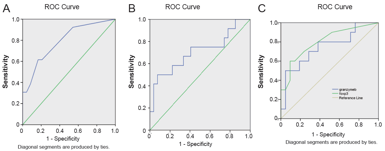
For comparison of the gross achievement of the biomarkers, distinctly of the pre-specified threshold levels, we tested the comprehensive degree of test performance using the AUROC. As shown in Figure 2A and Figure 2B, the AUC of FoxP3 mRNA expression levels were 0.792 with SE 0.078 (95% CI: 0.638–0.945; P ¼ 0.004) and serum granzyme B levels were 0.707 with SE 0.099 (95% CI: 0.512–0.902; P ¼ 0.041), respectively. It was as well detected that the co-evaluation AUC of FoxP3 mRNA expression levels and serum granzyme B levels were AUC ¼ 0.748 with SE 0.099 for granzyme B levels (P ¼ 0.028) and AUC ¼ 0.802 with SE 0.089 for FoxP3 mRNA expression levels (P ¼ 0.007) vs. AUC reference curve (AUC ¼ 0.5) (95% CI0.553-0.942, 0.629-0.976, respectively), similar to their individual AUC (Figure 2C). An optimal cutoff of FoxP3 mRNA expression levels was calculated at 0.1500 representing an equivalent specificity of 0.54 and sensitivity of 0.92, while the optimal cutoff of serum GZM-B levels was also statistically analyzed at 18.85 demonstrating an equivalent specificity of 0.85 and sensitivity of 0.92 (Table 2).
Multiple linear regression analysis showed that the hazard makers for AR in renal transplant recipients were FOXP3 mRNA levels (ß = 0.5, P = 0.003), serum granzyme B (ß=0.4, P=0.03) and serum creatinine level (ß=0.5, P=0.001) (Table 3).
|
|
|
|
|
|
|
DISCUSSION |
|---|
In this study, analysis of the impact of regulatory/cytotoxic pathways in acute rejection in pediatric renal transplant recipients has been performed. The faith is now arising that the balance of allo-aggressiveness to T cells protecting the graft is a specified predictor of allograft reaction—rejection or tolerance—post-transplantation (21). This balance is investigated by analyzing the regulatory/cytotoxic marker expression in T cells in peripheral blood from renal transplant recipients. It has been elucidated that T cell suppression of graft rejection is an active process that is implemented beyond secondary lymphoid tissue and includes the stable regulatory T cells present at the site of the tolerated allograft (18).
Post-transplantation of allogeneic organs, graft antigens causes activation of the circulating T cells in the transplant recipient which become sensitized, due to the existence of various histocompatibility antigens in both of donor and recipient. T cells can maturate and differentiate when entering the peripheral immune organs. Functional surface molecules are expressed on T cells which in turn secrete specified cytokines and differentiate into different T cell subsets (21). FOXP3 expressed in thymus and peripheral blood Tregs has a serious impact not only on the Tregs development but also their performance (22, 23). The present study declared that the expression levels of FOXP3 mRNA in children with AR were higher when compared with the expression levels of stabilized graft function; this result highlights a mechanism of negative feedback implicated in the immune system regulation. Our results suggested that Tregs together with the expression of FoxP3 as well as allogeneic antigens remaining in the transplant recipients are reproduced that do a regulatory function negatively to restrict anti-allograft immunity. It is also probable that the host anti-donor immune response during AR involves the activation of both the graft-destructive cytotoxic effector cells and the graft-protective Treg cells.
The level of FOXP3 messenger RNA expressed in peripheral blood mononuclear cells mirrors the condition of renal allografts and may be a noninvasive biomarker for the diagnosis of acute rejection. In our previous study, we reported that monitoring of circulating T cell activation marker i.e.CD25 in peripheral blood is useful for assessing the immune status of kidney transplant recipients during the early posttransplant period and associates significantly with the occurrence of AR in renal transplant pediatric recipients (24).
Previous researchers have reported that the expression levels of FOXP3 mRNA are associated with the concentration and type of the immuno-suppressive drugs utilized to prevent graft rejection (25). A low dose of tacrolimus given to kidney-transplant recipients may influence the number and function of Tregs in the peripheral blood (26). Also, recipients of renal-transplant age together with the post-transplantation time scale are correlated with the number of peripheral blood Tregs (27). In this study, differences elucidated between the group with clinically stabilized allografts and the AR group were not significantly different as regards to immunosuppressive regimens utilized and patient's age, so these results excluding the effect of these variables on FOXP3 mRNA expression.
Granzymes and perforin are the major influencing cytotoxic lymphocytes molecules (CTLs). Granzyme B is the utmost broadly searched granzyme promoting the death of cell where it stimulates the caspase-dependent and -independent pathways (28). The expression level of its gene in the peripheral blood was significantly high at the time of biopsy. Also, its relationship with perforin as molecular biomarkers of rejection is corroborated by researches demonstrating significantly higher levels of both of them in the urine of patients with AR, during the first 9 days and after the first 30 days post-transplantation 29. Histological researches have reported a plenty of granzyme B and perforin in numerous sorts of AR allografts (29).
There are different agents which can possibly bring down the specificity of the analysis based on gene expression of lymphocyte. Decreased bioavailability of cyclosporine is accompanied by the elevated expression of the gene in lymphocytes post-transplantation, but the elevated expression of granzyme B was demonstrated in patients with AR in spite of sufficient cyclosporine trough levels. A furthermore issue is the clinical infection in the late post-transplantation period, but prognosis of AR during the first 10 days might, in most of the patients, outdistance infection that accompanies the long-term immunosuppression, like CMV (We found that infectious complications such as CMV infection also did not lead to any significant changes in either of regulatory FoxP3 mRNA expression or cytotoxic GrzB levels.) Moreover, AR histological signs, seen in monitoring biopsies of patients free of clinical signs of AR (23), together with the reality that we did not have monitoring biopsy results for non- AR patients in the study, counsel that some samples in the study looked as false-positive may result from cases with ‘subclinical’ rejection. A prospectively randomized research was directed to detect that recovered function and survival of graft could be accomplished in patients with monitoring biopsies and management of subclinical rejection (25). Transcription of the gene in monitoring biopsy infiltrate was comparable phenotypically to that of infiltrate from clinical AR biopsies with the expression of granzyme B, but at a somewhat decreased level in subclinical when compared to clinical biopsy infiltrate (18). In our previous study, we found that FASL gene polymorphisms in peripheral blood may do an impotent function in AR. It could supply predictive investigation into the pathobiology of AR (30).
ROC curve detected that scaling FOXP3 mRNA may have a prospect as a decision-doing agent in clinical follow up to diagnose AR in a less invasive way. The AUC values are between 1.0 and 0.5, and the value is more near to 1, signalizing the preferable diagnosis is. Regarding accuracy, AUC in 0.5–0.7 has less reliability, 0.7–0.9 has confirmed reliability, and above 0.9 has a top. Also, on correlating AR to multiple risk factors by multiple linear regression analysis, we detected that serum FOXP3 was the most significant variable that was independently connected with AR. All these outcomes drive a hypothesis that FOXP3 might be not only a top marker to estimate the condition of immunity of renal transplant recipient but additionally a non-invasive tool for the diagnosis of renal allografts AR. Put together up- organizing of gene expression as a diagnostic standard for AR mostly elevated the specificity to detriment of sensitivity in rapprochement for the investigation of a single gene only.
The utility of novel immunosuppressive regimens in the latest decade has noticeably reduced the incidence of actual AR. We assume that contrariwise, the incidence of renal biopsy detecting restricted infiltrate like borderline changes or found out in the path of subclinical rejection will raise. Yet the query will be: “Are they deleterious or not”? We propose herein that the immune-monitoring of the ratio between the regulatory expression and a cytotoxic biomarker in the peripheral blood in pediatrics may be beneficial to investigate the immunologic tendency of this rejection.
|
|
CONCLUSION |
|---|
The study elucidated significantly increased expression of a regulatory and a cytotoxic marker, t FOXP3 expression /granzyme B levels the peripheral blood in the period of AR episodes, proposing that surveillance of the level of these markers could be utilized in the estimation of post-transplant children’s immune status that supplies worthy data for clinical management strategies. Though the moderate sample size in this study is considered a limitation, yet it could be suitable as this search was conducted on the pediatric population. Furthermore, these findings symbolize a single center experience of a racially homogeneous cohort and could not be applicable to other populations. Expression of FOXP3 mRNA and graft AR relationship need to be intensively searched and completed to resolve the relationship between graft AR and expression of this delegate biomarker of Treg cells and considering this marker as a valuable diagnostic agent for AR. These datums also emphasize how much substantial it will be for further studies of gene expression in transplantation to be done on extensive cohorts allowing for prospect confusing agents to be taken into consideration when assessing predictive, mechanistic or diagnostic biomarkers.
|
|
CONFLICT OF INTEREST |
|---|
The authors declare that no conflicting interests exist.
|
|
ACKNOWLEDGMENTS |
|---|
Our work was supported by the National Research Centre, Cairo, Egypt.
|
|
REFERENCES |
|---|