| ORIGINAL ARTICLE |
|
|
Department of urology, Military Hospital of instruction of Tunis, Tunisia
Corresponding Author: H. Ichaoui, Department of urology, Military Hospital of instruction of Tunis, Tunisia. E-mail: hamzaichaoui360@gmail.com.
| |
ABSTRACT |
| INTRODUCTION | |
|
|
OBSERVATION |
|
|
DISCUSSION |
|
|
CONCLUSION |
|
|
CONFLICTS OF INTEREST |
|
|
REFERENCES |
|
|
ABSTRACT
|
|---|
Condylomata acuminata are sexually transmitted papillomatous proliferative lesions caused by the Human Papilloma Virus (HPV) infection. Both sexes can be affected. In male patients, these lesions mostly occur on the glans, frenulum, prepuce and the balanopreputial sulcus. Urethral condyloma is relatively uncommon and it is frequently situated in the distal part of the urethra. We report the case of a 25-year-old male patient, single, whose medical history revealed that he had one unprotected sexual intercourse two years ago. He was presented in our department with a papillary lesion at the urethral meatus that was diagnosed as condyloma acuminatum which was confirmed later by performing a histopathological examination. A local excision of the lesion along with the use of podophyllotoxin as a local adjuvant therapy have led to its disappearance (no local or remote recurrence was seen 8 months later). Through this study, we aimed to discuss the different therapeutic approaches of urethral condylomata and mainly to assess the efficacy of podophyllotoxin as an adjuvant therapy after surgery.
KEY WORDS: condylomata acuminata; urethra; treatment; Human papillomavirus|
|
INTRODUCTION |
|---|
Condylomata acuminata (also variously known as papilloma or venereal warts) are secondary to a sexually transmitted infection (STI) caused by the Human Papillomavirus (HPV). Among the wide variety of HPV strains, the serotypes 6 and 11 are by far the most predominant types in this infection (1). The resulting lesions may be developped not only in the anal margin and anal canal but also in the genital area; in male, it affects the penile shaft, particularly the glans and prepuce, and the vulva, cervix and occasionally inside the vagina in female patients. The urethral and the bladder warts are relatively less common and the oral localization is practically exceptional (2). All of these possible sites of this infection justify a rigorous clinical examination of the perineum and therefore may require the cooperation between clinicians from multiple medical specialties: dermatologist, proctologist, urologist and gynecologist.
We report a case study of an urethral condylomata acuminata; the circumstances and the therapeutic approach were detailed in this study.
|
|
OBSERVATION |
|---|
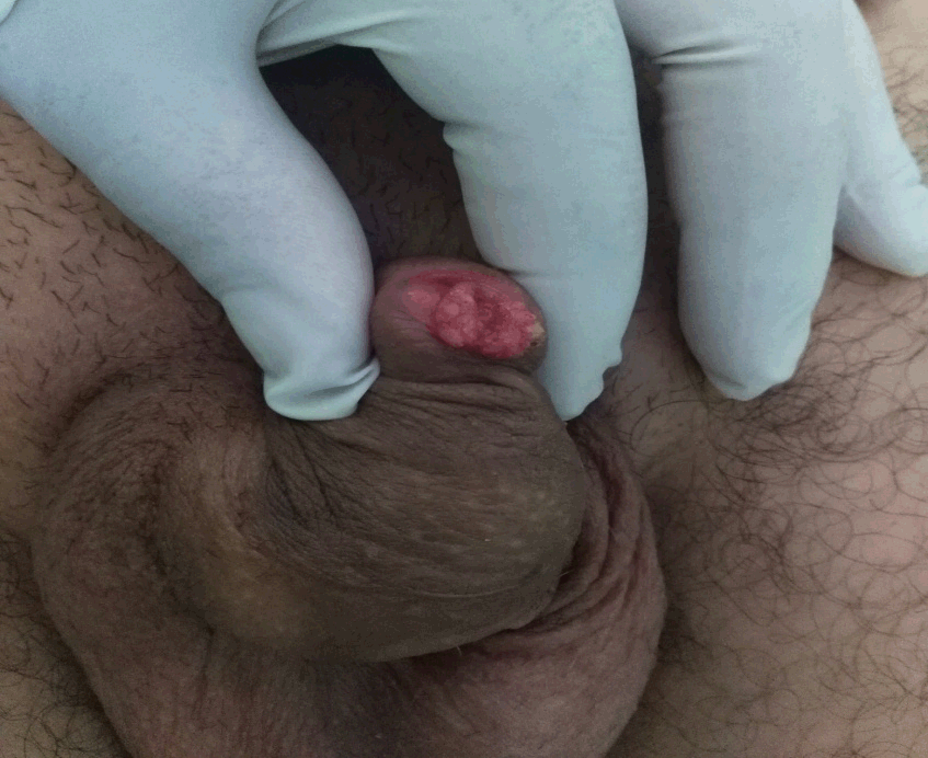
Our case was a 25-year-old male, single, he had one unprotected sexual intercourse two years ago. He attended us for tingling of the urethral meatus lasting for 3 months without any miction disorders or hematuria associated. Physical examination revealed a cauliflower-like tumor with budding and vegetating lesions located in the navicular fossa (Figure 1). It was diagnosed as condylomata acuminata and the cystoscopy did not reveal any further localizations. He was treated by a local excision of the lesion. The histopathological examination showed an exophytic proliferation made of an aconthotic papillomatous epithelium. Several koilocytes and dyskeratotic cells with parakeratosis and cytonuclear atypias have been noted. No microinfiltrant or infiltrating sites were observed.
HIV, syphilis, HBV and HCV serologic tests were negative in our patient.
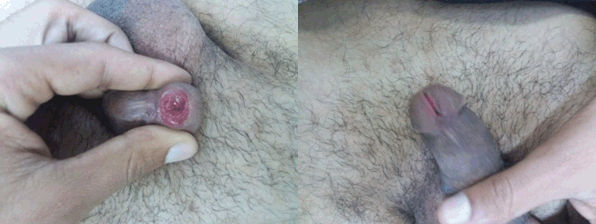
The excision of the condylomata associated with a local adjuvant therapy based on podophyllotoxin 0.5% (3 applications per week for 5 weeks) has led to the total eradication of the lesion (no recurrence was seen 8 months later) (Figure 2).
|
|
|
|
DISCUSSION |
|---|
Condylomata, also called papilloma, are growths similar to warts, they can be whitish, pinkish or grayish, or with serrated ridges (condylomata acuminata). Their size varies from a simple pinhead to vegetative formations of several centimeters. More rarely, it is flat lesions of small size at the limit of the visibility, whose diagnosis becomes more delicate (3).
The diagnosis of the condylomata is done by visual inspection, identifying them by the aspect of the lesions (4-6). Biopsy is only indicated in case of atypical lesions (7).
They are caused by several strains of HPV and are transmitted by skin-to-skin contact during sexual activity. They are therefore considered as a sexually transmitted infection (STI) (8). In more than 90% of cases, condylomata are caused by HPV type 6 and 11 which are weakly oncogenic (9).
In women, they can occur on the cervix, the vulva, inside the vagina or in the perianal area. In men, they can be located on the scrotum, the penis, the glans and under the prepuce or in the perianal region (10). The urethral and bladder localizations are much rarer and the oro-pharyngeal site is exceptional (2, 4, 11).
Microscopically, the CA is a complex papillary proliferation of a squamous epithelium, undulating and hyperplastic (acanthosis and papillomatosis) with an important hyperkeratosis often parakeratotic. This epithelium contains numerous koilocytes (vacuolized, clear cells with a central pycnotic nucleus surrounded by a clear halo) which are seen mainly in the superficial layers (5, 6, 12).
Although urethral condylomata are often asymptomatic, in other cases they can cause pain, tingling, miction disorders (5) and occasionally bleeding especially during the sexual intercourse (13).
The treatment of symptomatic CA is aimed at the attenuation of physical symptoms and cosmetic improvement. 40% to 60% of untreated condylomas will disappear spontaneously within 9 to 12 months (14, 15), but many patients experience psychological distress due to their presence and need an intervention to eradicate them (16).
Many therapeutics were used for the treatment of urethral warts. The aim of the treatment is the macroscopic destruction of lesions (4, 5) either by physical agents (electrocoagulation with electrocautery, cryotherapy, laser vaporisation, photodynamic therapy or scissors excision) or by chemical agents (podophyllotoxin, 5-fluorouracil or imiquimod) (4, 5, 17).
A mapping of the lesions made by cystoscopy is essential. The choice of the therapeutic method depends on the seat, size and number of lesions.
For unifocal meatal condylomata or of the distal urethra, scissors excision or destruction by liquid nitrogen are the most common methods. The laser did not decrease the recurrence rate and postoperative pain compared to other treatment methods (18). Photodynamic therapy with aminolevulinic acid can only be used in cases of single endocannal injury and its efficacy remains to be validated.
The destruction of urethral CA by the chemical agents (in local applications) is only indicated for the meatal lesions. They cannot be used on intra-ductal lesions. Podophyllotoxin such as fluorouracil and imiquimod may cause local secondary reactions like urethrostenosis, erosions and pain (5, 6, 12).
These topical agents are similar in terms of efficacy (17, 19). Their adverse effects are often reversible, moderate and fade after a few days of application of a topical corticosteroid.
The 'monotherapy' treatment of CA (by physical or chemical agents) is in fact of variable effectiveness and is often associated with a high recurrence rate (1, 17, 20, 21).
The combination of physical destruction of CA with topical application of the drug for accessible urethral lesions has been reported only rarely in the litterature (21-23). This combination appears to be associated with a lower recurrence rate with acceptable side effects.
We believe that combination therapy can be beneficial in decreasing the risk of recurrence of CA, and our patient’s case may be evidence of its effectiveness.
|
|
CONCLUSION |
|---|
Condylomata acuminata are often a benign condition even though the oncogenic role of HPV is real. It is the most common sexually transmitted disease. The recurrence after treatment is very frequent, sometimes desperate, requiring several destructions by physical or chemical agents. Therefore, we believe that the combination therapy associating a physical agent and a topical drug allows a better control of this disease while avoiding the repeated destruction of the lesions.
|
|
CONFLICTS OF INTEREST |
|---|
The authors declare that no conflicting interests exist.
|
|
REFERENCES |
|---|