| Original Articles |
|
|
1Key Laboratory of Environment and Genes Related to Diseases of Education Ministry, School of Medicine, Xi’an Jiaotong University, Xi’an, 710061, China
2Department of Obstetrics and Gynecology, Xijing Hospital, Fourth Military Medical University, Xi’an 710032, China
Corresponding author:
Tianbao Song Address:
Key Laboratory of Environment and Genes Related to Diseases of Education Ministry,
School of Medicine, Xi’an Jiaotong University, Xi’an,Shaanxi 710061, China
Tel:86-29-88069258 Fax:86-29-88390855
E-mail:ylhmqq@yahoo.com.cn
Running head Effect of ATZ on ovarian cancer cell
| |
ABSTRACT |
| INTRODUCTION | |
|
|
MATERIAL AND METHODS |
| RESULTS | |
|
|
DISSCUSSION |
|
|
REFERENCES |
|
|
ABSTRACT
|
|---|
Objective: To study the effect of 3`-azido-3`-deoxythymidine (AZT) on telomerase activity and the proliferation of ovarian cancer cells in vitro. Methods: Telomerase activity was detected by enzyme linked immunosorbent assay (ELISA) in treated and untreated HO-8910 cells by AZT. The detection of cell viability was performed with 3-(4,5-dimethylthiazol-2-yl)-2,5-Diphenyl tetrazolium bromide (MTT) assay and the ultrastructure of the cells was observed by electron microscopy. The apoptotic rate of the cells was measured by flow cytometry. Results: AZT significantly inhibited telomerase activity of HO-8910 cells, and the effect was both time- and dose-dependent. The HO-8910 cells treated at different concentrations of AZT showed a significant reduction of cell viability and morphological changes of apoptosis. The apoptotic peak was detected in the AZT treated cells and the apoptotic rate was 14.2%. Conclusion: AZT can effectively inhibit both telomerase activity and proliferation of human ovarian cancer HO-8910 cells in vitro, suggesting that AZT may be used in the clinic treatment of ovarian cancer.
KEY WORDS: 3`-azido-3`-deoxythymidine (AZT); reverse transcriptase inhibitor; telomerase; proliferation; ovarian carcinoma cell
|
|
INTRODUCTION |
|---|
Telomerase is a specialized reverse transcriptase (ribonucleoprotein polymerase) consisting of telomerase RNA and protein. Telomerase RNA with a short template element directs the synthesis of telemetric repeats at chromosome ends, maintains chromosomal stability, stabilizes telomere length, and leads to neoplasm occurrence and cell immortality [1, 2]. The telomerase activity was expressed in 85% of human cancers but not or seldom in normal somatic cells and benign tumors [3, 4]. Telomerase activation was thought to be an essential event for tumor proliferation [5, 6]. 3`-azido-3`-deoxythymidine (AZT) belongs to nucleoside analogs, which is currently used in the treatment of acquired immunodeficiency syndrome (AIDS). The main effect of AZT is to inhibit the activity of reverse transcriptase and synthesis of virus [7, 8]. In recent years, researchers have found that AZT can inhibit many enzyme activities of cells in vitro, especially the telomerase activity. AZT can also inhibit reverse transcription process, activity of reverse transcriptase, telomerase activity and telomere expanding. AZT can also inhibit cell division and telomere shortening. Thus cell stability is disrupted, and cell proliferation and growth are inhibited [9, 10].
Ovarian cancer is the most common malignant tumor of women, with high mortality
and poor prognosis. Although advances in ovarian cancer treatment with
cytoreductive surgery and chemotherapy have improved survival rates in the last
decade, the prognosis remains poor in patients with ovarian carcinoma because of
ineffective diagnosis and treatment. Therefore, it is essential to explore novel
forms of diagnosis and treatment. Numerous studies have demonstrated that
telomerase is highly expressed in ovarian cancer [11, 12, 13]. In this study,
the inhibitory effect of AZT on telomerase activity and proliferation of ovarian
cancer cells in vitro were examined and the possibility of AZT for clinical
therapy of ovarian cancer was explored.
|
|
MATERIAL AND METHODS
|
|---|
Regents
AZT was purchased from Sigma (St. Louis, MO, USA), telomerase ploymerase chain
reaction-enzyme linked immunosorbent assay (PCR-ELISA) Kit from Boehringer
Manheim (Germany), RPMI-1640 from Hyklong Company (USA), and trypsin from DIFCO
Company (USA). Penicillin and streptomycin were the products of Pharmaceutical
Factory of Haerbin, (China), fetal calf serum was obtained from Zhejiang
Evergreen Company (China), 3-(4,5-dimethylthiazol-2-yl)-2,5-Diphenyl tetrazolium
bromide (MTT) from Huamei Biological Company (China), and dimethyl sulfoxide
from Jinshan Chemical Plant (Shanghai, China).
Cell line
Ovarian cancer cell line HO-8910 was developed in the laboratory of the
Department of Obstetrics and Gynecology, Xijing Hospital, China. HO-8910 cells
were cultured in RPMI 1640 supplemented with 100mg/ml streptomycin, 100mg/ml
penicillin and 10% fetal calf serum. The cells were incubated at 37℃ in the
presence of 5% CO2. Logarithmically growing cells were used for subsequent
experiments.
Telomerase activity assay
HO-8910 cells were randomly divided into experimental and the control groups.
Cells in the experimental group were maintained in culture medium with
supplemented with AZT at he concentrations of 0.5 mM,0.8 mM,1.0 mM and 1.5 mM
for 24h,48h and 72h, respectively. The same culture medium without AZT was used
for the cells in the control group. Cells were collected at different time
points by centrifugation at 2000g for 10 min at 4℃. The pelleted cells were
resuspended in 200μL lysis reagent and incubated on ice for 30 min followed by
centrifugation at 12,000g for 25 min at 4℃. In the control group, HO-8910 cells
were treated for 10 min at 65℃. The telomerase activity of HO-8910 cells was
measured using a telomerase PCR-ELISA Kit .
MTT assay on suppression of tumor cell growth
HO-8910 cells were cultured in RPMI 1640 containing 10% fetal calf serum, in
which the cultured cells reached over 90% confluence in 96 well plates.
Different concentrations of AZT (0.05 mM, 0.1 mM, 0.2 mM, 0.5 mM, 0.8 mM, 1.0 mM,
1.2 mM, 1.25 mM, 1.6 mM, 2.0 mM) were added to the cells in the experimental
group while equal volumes of culture medium were added to the cells in the
control group. After 24, 48 and 72h, 20μL of MTT(5mg/mL)was added to each well
and the cells were incubated for another 4h at 37℃. The supernatant was
carefully removed, and 150μL of dimethyl sulfoxide was added into each well and
shaken for 10 min. Absorbance (A) of the samples was measured at 490 nm by an
enzyme linked immunoassay meter. The inhibitory rate of tumor cells = (1- AAZT/Acontrol)
×100%.
Ultrastructure of tumor cells
HO-8910 cells were cultured in 100 mL flasks in RPMI 1640 containing 10% fetal
calf serum. When the cells reached over 90% confluence, AZT was added to the
cells in the experimental group at the final concentration of 0.8 mM or 1.2 mM,
and equal volume of culture medium was added to the cells in the control group.
Subsequently, cells in both groups were incubated for another 72h., after which
cells were transferred to a fresh tube and collected by centrifugation at 2000g
for 10 min. 2 mL of 20g/L glutaraldehyde was then added and the cells were fixed
for 2h. After cells were embedded and ultrathin sections were cut, the
ultrastructure of the cells were observed by transmission electron microscope.
Cell cycle definition
HO-8910 cells(5×10⁵)in the experimental group were added to culture medium
containing 0.8 mM or 1.2 mM AZT and the cells in the control group were added to
culture medium without AZT. After incubation for 72h, the cells in both groups
were collected by centrifugation at 1000g for 10 min and the pellet was
carefully washed twice with 0.01M phosphate buffered saline (pH 7.3). Then 1mL
of 70% ethanol was added to the collected cells and the cells were kept at 4℃
for further processing. After washed twice with 0.01M phosphate buffered saline,
the cells were stained with 300μL of propidium iodide for 30 min. The cell cycle
was detected by flow cytometer.
Statistical analysis
The SPSS 12.0 software was used to determine the statistical significance of the
differences between the two groups. The result was examined by the Repeated
Measure ANOVA. P value less than 0.05 was considered significant.
|
|
RESULTS |
|---|
Inhibition of telomerase activity of HO-8910 cells by AZT
Telomerase activity of HO-8910 cells was inhibited by AZT at 0.5 mM for
24h. The inhibitory effect was significantly increased with the
elevation of drug concentration and elongation of exposure time.
(P<0.05, Table 1).
|
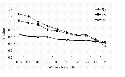
Suppression of cell growth with MTT assay
The inhibition on HO-8910 cell growth of AZT at different concentrations for
24h, 48h and 72h, respectively, was detected with MTT assay. The results showed
that lower concentrations of AZT had a slight inhibitory effect on HO-8910
cells. With the increase of AZT concentration, the A value was decreased and the
inhibitory rate was increased and the effect was dose-dependent. Similarly, with
the elongation of acting time, the inhibitory rate was also increased in a
time-dependent manner. The minimum inhibitory effect appeared with 0.8mM at 24h,
with 0.5mM AZT at 48h , and with 0.1mM AZT at 72h (Fig. 1, Table 2).
|
|
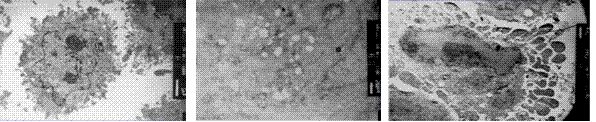
Ultrastructural changes of HO-8910 cells
The cells in the control group were observed to be polygon- or anomalous-shaped
with various sizes by electron microscope. There were lots of long and thin
microvilli on cellular surface (Fig. 2A). These cells had well-developed
organelles, including mitochondria, rough endoplasmic reticulum, lysosomes and
free ribosomes, etc. The nucleus was large with many karyokinesis, and abnormal
nucleus mitosis was observed. After treated with AZT at 0.8 mM for 72h, some
HO-8910 cells became swollen with dilated endoplasmic reticulum and fewer
mitochondria appeared when compared with the cells in the control group (Fig.
2B). After treated with AZT at 1.2 mM for 72h,the microvilli of the cells were
decreased in number and plenty of bubble-shaped convex appeared on cell surface.
The chromatin was condensed and aggregated against the nuclear membrane, the
structure of which was intact (Fig. 2C).
|
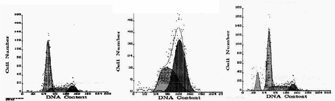
Changes of cell cycle
The cells treated with AZT at various concentrations had cell cycle profiles
different from those of the cells in the control group. After exposure to 0.8 mM
AZT for 72h, the percentage of the cells in the phase G1 dropped from 57.6% (in
control group) to 1.1%,the percentage of the phase G2/M cells rose from 15.5% to
57.6%, and the percentage of the phase S cells rose from 26.9% to 41.2%. When
AZT was used at the concentration of 1.2 mM, the percentage of the phase G1cells
rose to 67.7%,the phase G2 cells fell to 12.2%,and the phase S cells dropped to
20.1%. The apoptosis cusp was observed and the apoptotic rate was 14.2% in the
group of 1.2 mM AZT (Fig. 3).
|
| |
|---|
AZT is a nucleoside analog able to inhibit reverse transcriptase by competing
for deoxynucleotides, resulting in termination of chain elongation [1]. AZT was
originally used to treat AIDS. AZT not only inhibits certain enzyme activities
of cells but also induces apoptosis of lymphoma correlated with AIDS[14, 15,
16]. It is reported that AZT can inhibit telomerase activity and proliferation
of mammary cancer cells, cervix cancer cells, and other cancer cells in
vitro[17, 18].
In our study, we observed the effects of AZT at different concentrations on
HO-8910 cells of ovarian cancer and their results at different time points. Our
findings showed that AZT effectively inhibited telomerase activity and
proliferation of human ovarian cancer HO-8910 cells in vitro, and that the
viability of cancer cells had a significant reduction. At the same time points,
the inhibitory effect of AZT was increased with the elevation of drug
concentration; at the same drug concentration, its inhibitory effect was
significantly increased with elongation of time. The inhibitory effect was both
time- and dose-dependent. The notable cytotoxic effect of AZT on HO-8910 cells
at 0.1 mM was observed 72h after drug treatment and at 0.8 mM 24h after AZT
administration. The results indicated that drug concentration and exposure time
of AZT were important for the suppressed growth of ovarian cancer HO-8910 cells.
Cellular swelling, endoplasmic reticulum expanding and mitochondrium were
observed to decrease in number by electron microscopy. The chromatin of cells
was condensed and peripherally located. Flow cytometry detection showed that the
cells treated with the drug at different concentrations had different cell
cycles. The cells treated at low concentrations of AZT (0.8 mM) for 72h were
gathered at the phase G2/M. When the cells were treated with 1.2 mM of AZT for
72h the cells at the phase G1 increased and the cells at the phase S decreased.
The apoptotic peak was detected by flow cytometry and the apoptotic rate was
14.2%. The growth suppression of cells might be related to apoptosis.
Nevertheless, it is still unknown whether AZT itself or apoptosis inhibiting
genes induce the apoptosis, and the mechanism of cell apoptosis needs further
investigation.
We also found that some cells survived at the 2.0 mM of AZT concentration due to
their resistance to AZT. The development of these cells and their relation to
drug resistance of tumor cells require further study.
Some researchers reported that the effect of tumor chemotherapy was more obvious
when AZT was combined with drugs for chemotherapy. AZT may act as a synergist of
drug for chemotherapy [19, 20]. In our previous research we studied the effect
of AZT combined with the drugs of chemotherapy on the growth of human ovarian
cancer line HO-8910. The inhibitory effect on cancer cells of AZT combined with
adriamycin or carboplatin was stronger than that of each drug singly used [21].
Chemotherapy is an essential therapeutic approach in the treatment of ovarian
cancer. The results from our research showed that AZT had some significantly
inhibitory effects on telomerase activity and proliferation of HO-8910 cell line
of ovarian cancer cells. The ideal targeting strategy for tumor therapy should
focus on some essential components present in tumor cells but not in normal
cells.Telomerase is an essential condition of cell immortalization and an
important factor of tumor development. Theoretically, the inhibition of
telomerase activity by AZT may become a new treatment target because AZT can
directly inhibit telomerase, which is absent in normal somatic cells. AZT can
inhibit the growth of tumor cells with little injury to most normal cells by
inhibiting telomerase activity. Moreover, it may also increase the specific
effect and reduce the side effects of chemotherapy.
Since most tumor cells have telomerase activity, treating cancers by inhibiting
telomerase activity is of great prospect [22]. Therefore, the application of AZT
might provide a novel approach to the targeted treatment of ovarian cancer and
other cancers. However, the mechanism by which AZT inhibits the cell growth and
interrupts the cell cycle of ovarian cancer needs further investigation.
|
|
REFERENCES
|
|---|