| REVIEW ARTICLE |
|
|
1 SCB Dental College and Hospital, Cuttack, Odisha, India;
2 Associate Professor, Department of Dentistry, PRM Medical College and Hospital Baripada, Mayurbhanj, Odisha, India;
3 Assistant Professor, Department of Dentistry, PRM Medical College and Hospital Baripada, Mayurbhanj, Odisha, India;
4 Dental Surgeon, District HQ Hospital, Department of Dentistry, PRM Medical College and Hospital, Baripada, Mayurbhanj, Odisha, India;
5 Senior Resident Department of Dentistry, PRM Medical College and Hospital Baripada, Mayurbhanj, Odisha, India;
6 Junior resident Department of Dentistry, PRM Medical College and Hospital Baripada, Mayurbhanj, Odisha, India
Corresponding Author: Tribikram Debata, SCB Dental College and Hospital, Cuttack, Odisha, India. E-mail: tdebata987@gmail.com.
Running title: EFFECTS OF VITAMIN D DEFICIENCY ON DENTAL HEALTH: A SYSTEMATIC ANALYSIS
| |
ABSTRACT |
| INTRODUCTION | |
|
|
MATERIALS AND METHODS |
|
|
RESULT AND DISCUSSION |
|
|
CONCLUSION |
|
|
DECLARATION OF PATIENT CONSENT |
|
|
ACKNOWLEDGEMENTS |
|
|
FINANCIAL SUPPORT AND SPONSORSHIP |
|
|
CONFLICTS OF INTEREST |
|
|
REFERENCES |
|
|
ABSTRACT
|
|---|
Vitamin D is an important micronutrient for human health that is stored in body fat. Vitamin D deficiency is a health problem all over the world. The purpose of this study is to provide a concise summary of the current literature on the topic of vitamin D status in India with the ultimate goal of better-comprehending vitamin D deficiency and its effects on dental and oral health. These studies have found that vitamin D deficiency is quite common with estimates ranging from 40 to 99 %. All age groups and high-risk populations were affected. Vitamin D stimulates tooth enamel mineralization by binding to the vitamin D receptor (VDR), which is expressed in a variety of cell types including those that makeup teeth and bones. The public and medical professionals both need to be made more aware of vitamin D's significance and the negative effects of insufficiency. The average Indian diet lacks the vitamin D needed by an adult body. This analysis was conducted to determine how well the public is informed about the role that vitamin D plays in maintaining good oral health. Increasing the amount of vitamin D in the body can reduce the risk of dental caries, periodontitis, and oral cancer in humans.
KEY WORDS: Vitamin D; Deficiency; Oral Health|
|
INTRODUCTION |
|---|
India has a high number of dental schools per capita but the country still facing oral health problems. Oral cancer and gum disease are major public health issues in the United States (1). To make national policy or allocate resources, India needs more comprehensive data on the prevalence of oral diseases due to the country’s size and population (2). Also, there are 1.21 billion people in the world and 68.84% of them live in rural areas. As a result, there is not much importance on research being done, and policy being made regarding the infrastructure and resources for oral health (3).
Dental caries, also referred to as tooth decay, is the most prevalent oral disease condition. As a result of bacteria adhering to the tooth’s surface and converting carbohydrates into acid which demineralized and dissolved the tooth structure (3). According to research, vitamin D supplements reduce dental caries but more investigation is required to established the association of the caries and vitamin D. Some reviews of the literature explained vitamin D dissolves in fat-like sterols and works as a hormone (2). The active form of vitamin D is calcitriol, which is produced in kidney. Vitamin D can be consumed or added to food and it can also be produced by the skin when it is exposed to UV light It can also dissolve fat (3).
Oral health is paying more attention to Vitamin D levels. VD deficit (VDD) is related to oral health issues during childhood and adulthood. Also reduced VD synthesis may accelerate certain illnesses. Severe VDD can cause dentin and enamel abnormalities in children. These defects may increase the risk of dental caries (6).
Oral health issues in India and America
General health is closely related to oral health. Despite having the most dental schools worldwide, India nevertheless has a variety of oral health problems that are of concern. India lacks the baseline data necessary to deter- mine the precise prevalence of oral disorders, which is a prerequisite for developing national policies or allocating resources. India is a sizable country with numerous nations within it, and there is a wide range of dietary and behavioral customs. With 1.21 billion people and 68.84% of them living in villages, the plans must be tailored to the population segment. Lack of enthusiasm for research and oral health policy, a low level of awareness, stretched and uneven infrastructure and limited resources are some of the main issues that need to be addressed (4).
American Oral Health: Progress and Challenges demonstrate the advancements made and the issues still present since the publication of the Oral Health in America study in 2000.3 Through its state-managed Medicaid programs, the Children’s Health Insurance Programme (CHIP), and other market-place initiatives approved under the Afford- able Care Act, the USA has increased dental coverage over the past 20 years, leading to today’s nearly universal dental coverage for children. This development demonstrates how multilevel efforts in the execution of health policy initiatives can affect the outcomes for children’s oral health, particularly those living in poverty (5, 6) (Fig. 1).
To conduct a comprehensive search of the dental literature published between 1980 and 2022, we used electronic databases including PubMed, the database of the United States National Library of Medicine, and Thomson Reuters Web of Knowledge. The search terms included both MeSH (Medical Subject Headings) and free text terms, such as [periodontitis OR periodontal disease] AND [vita- min D OR calcitriol] AND [vitamin D deficiency].
|
|
MATERIALS AND METHODS |
|---|
Search Strategy
A comprehensive search approach was built based on keywords and MeSH terminology relevant to ‘Vitamin D Deficiency’ and ‘Oral Health’. The thorough search was conducted using the search terms “Vitamin D Deficiency” and “Oral Health”, “metabolites” and “Dental Caries” “Etiology” and “Vitamin D impact on oral health”, and “Role of vitamin D” and “vitamin D receptor (VDR) in oral cancer”. These were combined with Boolean opera- tors “AND/OR” and the search strategy was customized to each database.
Three online search engines – Medline (PubMed), Web of Science, and Scopus were independently searched by three authors to find qualifying studies. The review of the retrieved papers was performed in three steps, including title screening, abstract and full-text screening against inclusion and exclusion criteria. Two authors separately re- viewed the articles. Studies that did not meet the inclusion requirements were excluded. Following the title and abstract screening, the potentially relevant studies were evaluated and assessed for eligibility by way of full-text screening by three reviewers. Disagreements between authors have been settled by discussion and mutual consensus.
Selection Criteria
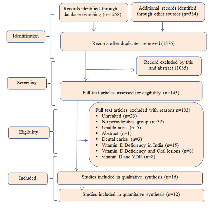
We only included the studies that provided estimates of the ambient Impact of Vitamin D Deficiency on Oral Health and all epidemiological studies including original articles, cross-sectional studies, cohort studies, longitudinal studies, case-cross-over studies, and intervention studies compare with review articles. Duplicate articles were removed. Editorials, review articles, case reports, studies on smoking tobacco, the effect of Oral Health, and studies focused on behavior only (without Oral Health) were excluded from the review Fig. 2.
The role of vitamin D in modern dental practice is not well understood and more research is needed. The purpose of this article was to show the effects of vitamin D deficiency on oral health.
|
|
|
|
RESULT AND DISCUSSION |
|---|
Dental caries and their origin
Dental caries is also called tooth decay. This is a disease of the hard tissues of the teeth that leads to irreversible demineralization of the inorganic part of the tooth and destruction of the organic part of the tooth which can cause cavitation (7).
Dental caries warning signs
The symptoms of dental caries can range from a chalky or pigmented enamel to cavitation.
Symptoms
Caries may be characterized by the experience of pain, and problems with eating, chewing, smiling, and communication due to missing, discolored, or damaged teeth (8).
Cause
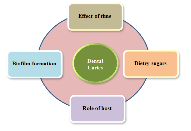
When bacteria ferment sugars in the diet, they produce acidic by-products that can cause localized destruction of soft dental tissues, this condition is known as dental caries. The ecological imbalance between tooth minerals and oral biofilms (plaque) leads to the slow, chronic progression of the disease (9, 10). In the biofilm, microbial activity causes pH changes in the plaque. This occurs because of the combined effects of bacterial acid production and the buffering action of saliva and the tooth structure around it. Thus, the tooth’s surface is in a state of dynamic equilibrium with its immediate environment. When the pH drops below a certain threshold, enamel, dentine, and cementum lose minerals and are demineralized conversely, when the pH rises above this threshold, these tissues gain minerals and are remineralized (11). Demineralization and remineralization occur repeatedly throughout the day. If left untreated, this process can cause caries lesions but with treatment, it can also be reversed (12). Caries, or cavity, in teeth, are typically brought on by bacteria living on the tooth’s surface. When bacteria accumulate on a tooth’s surface, they produce a sticky film. At the outset of caries, the enamel is demineralized due to the action of bacteria that secrete acids that do this. When enamel breaks down, it becomes thin and see-through. This lets bacteria get deeper into the tooth and remove minerals from the dentin which makes cavities (Fig. 3).
Vitamin D
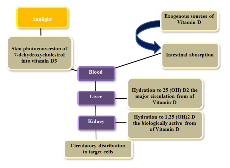
Vitamin D is a vitamin that works with fat. Vitamin D is sometimes referred to as the “sunshine vitamin”. Vitamin D functions as a prohormone. When the skin is exposed to sunlight, 7-dehydrocholesterol is changed into cholecalciferol which is then hydroxylated at the 25th position to make 25-hydroxycholecalciferol which is the main form of vitamin D that the body stores and uses. Calcitriol is the form of vitamin D that works in the body (13) (Fig. 4).
Magnitude of Vitamin D Deficiency in India
Vitamin D deficiency is considered to be common in countries with and without adequate sunlight. Even now, it is the dietary deficit that receives the least attention and the fewest diagnoses worldwide (14, 15). These studies used varying deficient cut-offs since there is no universally accepted method for defining vitamin D status. Most of these studies defined vitamin D insufficiency as a blood 25(OH) D level below 20 ng/ml. For the past ten years, research conducted in Indian communities using seemingly healthy controls has indicated a prevalence anywhere from 50% to 94%. However, one study only found a prevalence of 34.5%, which may be attributable to a too-low threshold. The scope of the issue is reflected by this research’s inclusion of people of varying ages (16, 26) in Table 1. Vitamin D deficiency is extremely common, with hospital studies estimating it to be as high as 99 % (27, 28, 49). Research on the link between not getting enough vitamin D and certain diseases was left out in Table 2.
Kadam et al. (2011) conducted research on premenstrual females (n=214) in a Pune school. Overall, vitamin D levels were found to be 34.2% (50). Another school-based study was conducted in 2017 by Kapil et al. on 1222 students aged 6–18 from the Kangra and Kullu districts of Himachal Pradesh, and they found a prevalence of 80% (17). In both investigations, Vitamin D deficiency prevalence was reported in both ways using the criteria established by the United States Endocrine Society (51) (Table 1 and Table 2).
Causes of Vitamin D Deficiency
Deficiency of vitamin D is common in India as seen in the tables above. Vitamin D deficiency occurs in hu- mans for a variety of reasons including poor dietary intake and illnesses affecting the liver, kidneys, and skin, this is due to the rise of the indoor lifestyle, which blocks off too much sunshine. As a result of progress, this is more common among city dwellers.
Role of Vitamin D and its effects on oral health
Eating a healthy and balanced diet can improve your overall health in addition to the benefits it affords your mouth (54). The relationship between vitamins and oral health has not been thoroughly researched or developed. Vitamins speed up the basic metabolic processes of the body which are important for cell health, growth, and development (55).
Magnesium, calcium, and phosphorus are essential structural components of teeth. It should be consumed in sufficient amounts through food. Teeth are strengthened by the interactions between minerals such as calcium, magnesium, and phosphorus. In particular, vitamin D is connected with calcium, magnesium, and zinc (54). Vitamin D has been shown to reduce the risk of caries in many ways.
Childhood cavities and temporomandibular joint dysfunction afflict kids everywhere. Both enamel and dentin undergo modifications in VDD-affected children. Because of this, vitamin D affects the growth of primary teeth and is a key part of making enamel and dentin, the hard parts of teeth (57). Vitamin D is crucial in the development of teeth (56, 57). Vitamin D stimulates enamel mineralization by binding to the vitamin D receptor (VDR), which is present in cells lining teeth and bones. A majority of the genes linked to ameloblasts and odontoblasts differentiation are controlled by vitamin D receptors (56). Dentin which contains structural gene products including calcium-binding proteins and other extracellular matrix proteins is prompted to develop in response to VDR activation. The VDR gene which encodes the protein is located on chromosome 12q13.11 and is highly polymorphic (58).
The VDR gene plays an important role in tooth development which notably in the mineralization of dentin and enamel by modulating the biological roles of main vitamin D metabolites. As a result, VDD can lead to deficits in enamel development such as enamel hypoplasia. Vitamin D and VDR have been shown at the molecular level to influence tooth germ development, provide control of enamel and dentin structure and maturity, and organize the stages of dental crown growth (54, 56).
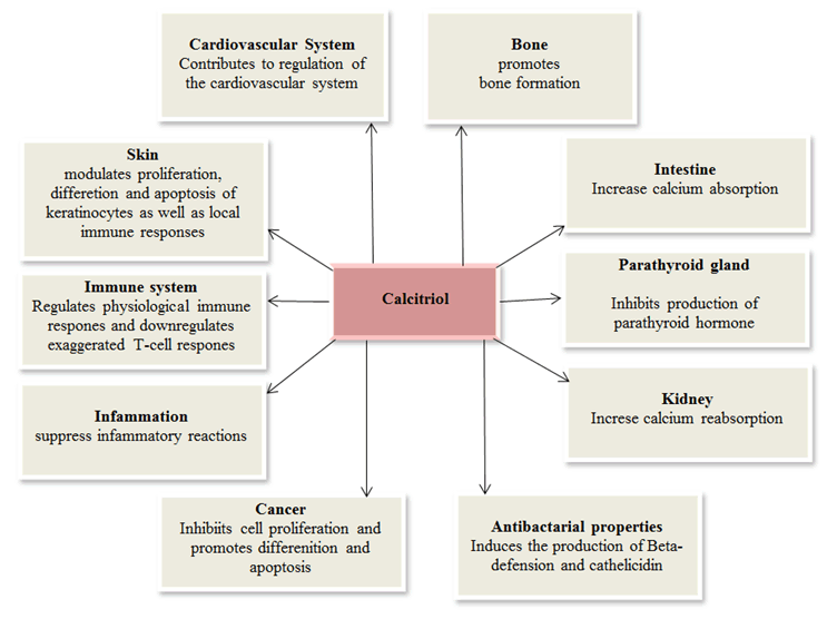
Vitamin D also regulates and fine-tunes both the innate and adaptive immune systems. Vitamin D has an important immunological function in coordinating the formation of antimicrobial peptides including defensins and cathelicidin (LL-37) which protect against a wide variety of infections, including oral bacteria (56). Vitamin D controls how much cathelicidin (LL-37 or hCAP-18), an antimicrobial peptide that fights endotoxins, is made in the body (59).
Absorption issues can lead to a lack of minerals which can increase the risk of bleeding, bone resorption, and pre- mature tooth loss (54). When a person has a complete set of teeth that are evenly spaced, they can chew food more efficiently and maximize nutritional absorption. When teeth are lost at a young age, it can have a domino effect on a per- son’s nutritional condition and diet and lead to a vitamin shortage. Throughout development, nutritional status and vitamin deficiency have a significant impact on the hard tissues of the teeth. Child malnutrition has been linked to the development of cavities and enamel hypoplasia during the primary dentition stage (55). Mineral deficiencies have been linked to tooth eruption delays, bleeding gums, destructive patterns in alveolar bone, periodontal disease, and underdeveloped enamel or dentin (55, 56).
In addition to minerals, vitamin D is also helpful in warding off damage to teeth and gums. Maintaining a healthy calcium-phosphate balance is essential for proper bone development and vitamin D plays a role in this process. Furthermore, it serves a crucial purpose by reducing inflammation. It has been observed that adequate vitamin D levels can halt the development of caries, lower the rate at which carries occur and prevent enamel loss (54, 56).
The acid created by bacterial fermentation of sweet food remains on the teeth surface that are not brushed away after eating. This contributes to the breakdown of tooth-hard tissues. More recent research, however, suggests that exposure to UV-B rays and vitamin D supplementation may help mitigate dental caries (54, 56). One of the most common chronic diseases that can harm an individual’s health is early childhood caries (ECC) is defined as the presence of one or more caries, missing (due to caries) or filled (DMFT: decay, missing, or filled) tooth surfaces in any primary teeth in children under the age of six (56).
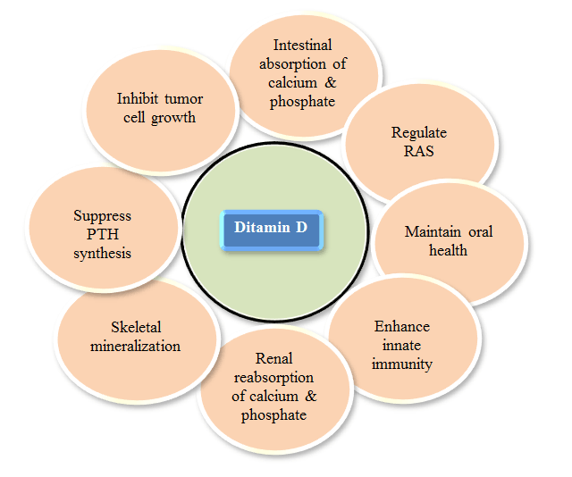
Vitamin D is a hormone that helps the body digest and absorb calcium, magnesium, and phosphorus which are all important for healthy bones and teeth. In addition, one of the dental treatments can be enhanced by coating the implant surfaces with vitamin D. This promotes osteointegration. Also, using vitamin D3 speeds up the movement of teeth in orthodontics and people who take vitamin D and bisphosphonate drugs can still get orthodontic care (56) (Fig. 5).
Vitamin D Deficiency and Oral lesions
Vitamin D may play a role in the initiation and progression of some oral malignancies. VDD is more common in patients with oral cancer (60). VDD increased the incidence of esophageal, oral, and pharyngeal malignancies in heavy smokers and alcoholics (61). VDR expression was higher in premalignant lesions and oral cancer and vitamin D administration reduced therapy-related toxicities in advanced stage of oral malignancies which is leading to decreased morbidity and a greater quality of life (62). Future research should elucidate how VDD affects oral cancer development and anticancer drug effectiveness.
The function of VDD in ONJ is widely studied but vitamin D status in ONJ patients is unknown (63-65). ONJ is defined as increasing jawbone loss in a patient consistently exposed to anti-absorption or anti-angiogenic drugs with no history of mandibular radiation or metastatic jaw illness. Vitamin D levels in ONJ cases have been studied due to their function in bone mineralization (64-70). Recent evidence suggests that VDD is not a risk factor for ONJ events while other studies suggest a potential role of VDD (64-65, 69, 70). Future research should concentrate on the impact of baseline VDD and Vitamin D supplementation on ONJ as well as the development of 25(OH)D level standards for patients at high risk of ONJ events.
Vitamin D Deficiency in Tooth Mineralization and Caries
Enamel, dentin, and cementum make up the teeth which are mineralized organs encased in alveolar bone. Failures in tooth mineralization are analogous to bone tissue failures if mineral metabolism is disrupted. Uncontrolled levels of vitamin D have been linked to rachitic teeth which are weak and prone to cavities because they don’t have enough mineralization to protect them (71, 72). There is much discussion elsewhere about the processes by which VDD impairs tooth mineralization (71, 72). The primary biological reason is the association between severe VDD (10 ng/mL) and hypocalcemia, hypophosphatemia, and secondary hyperparathyroidism (caused by hypocalcemia) (73, 74). Increased bone turnover causes increased blood Ca2+ and decreased serum Pi due to hyperparathyroidism which in turn enhances intestinal absorption of Ca2+ and renal synthesis of 1,25-dihydroxyvitamin D (1,25 [OH] 2D) (73, 74). After that, the original hypophosphatemia worsens dramatically. Lastly, mineralization problems happen when there aren’t enough Ca2+ and phosphate ions in tooth cells to keep vitamin D signaling pathways open (71).
Apart from its involvement in mineralization equilibrium, circulatory vitamin D can activate a signaling cascade via vitamin D receptors (VDR). The vitamin D response element (VDRE) has a role in gene expression and the vitamin D receptor (VDR) is a ligand-activated transcription factor that regulates this process (75). Some of these responsive genes have a role in skeletal muscle, detoxification, energy metabolism, immunological response, and bone and mineral metabolism (75-79). Vitamin D helps dentin and enamel grow by making structural gene products like calcium-binding proteins and extracellular matrix proteins (like enamels, amelogenins, dentin sialoglycoproteins, and dentin phosphoproteins) more likely to be made (71, 79).
Vitamin D Deficiency and Orthodontic tooth movement
Facial micro- and macro-aesthetics as well as the grin are becoming increasingly important to both young and old people (80, 81). For this reason, orthodontic care is more common. The mechanical stimuli necessary for tooth movement are applied in a controlled manner to 1) bone resorption at the pressure site (through osteoclastic activity) and 2) bone synthesis at the tension site (by osteoblastic action) (82–84). These two procedures when combined with mechanical, chemical, or electrical stimulation could hasten the movement of teeth (85-88). In terms of chemical considerations, vitamin D may have an important role in tooth mobility during orthodontic treatment. Although the majority of the research is based on observations of animals there is mounting evidence that shows local injection of vitamin D speeds up tooth movement (88–90). Despite this, VDD slows down tooth movement in animal models, which might induce treatment delays or difficulties (88-90).
More research needs on humans to find out the clinical impotence of VDD and its important effect on teeth movement. If vitamin D supplementation is used at the time of orthodontic treatment for example in cases of hypovitaminosis, it can improve the coupling formation and resorption in alveolar bone remodeling.
The effect of vitamin D and VDR on oral cancer
Oral cancer is a malignant neoplasia of the lips and oral cavity. More than 90% of all oral malignancies (92) are oral squamous cell carcinomas (OSCC). OSCC incidence rates are rising in several parts of the world in recent years. Multiple studies have found that using tobacco products and drinking alcohol together raises the risk of mouth cancer by about 80%. Human papillomavirus (HPV) infection of the mouth is a known risk factor for oral, pharyngeal, and throat cancer (93). When done after an oral cancer diagnosis, removing major risk factors improves prognosis and lowers recurrence risk (60).
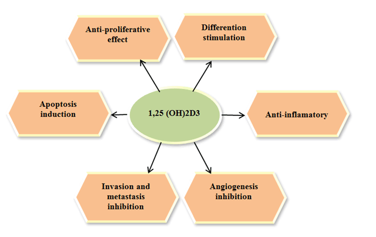
As a multi-step process, OSCC growth impacts critical biological processes linked to tumor genesis and progression. Multiple molecular alterations, both foreign and endogenous, have been linked to carcinogenesis. In vitro and in vivo investigations have shown that vitamin D (calcitriol) has an anti-neoplastic effect against a range of cancer-associated abnormalities including head and neck cancer and especially OSCC (94). Furthermore, it can enhance apoptotic induction in OSCC cells and influence cytostatic chemotherapy. It makes sense to look into the relationship between blood vitamin D levels and VDR to better target supportive care for patients with precancerous lesions and OSCC (60). Despite several in vitro and in vivo studies demonstrating vitamin D anticancer effects. Recent evidence shows that these effects are regulated by factors outside of vitamin D itself. Better VDD may slow the growth and spread of tumors but more research is needed to find out exactly what role the vitamin D system (ligand and receptor) plays in the development of oral cancer (Fig. 6).
Effect of Vitamin D on Periodontal pathology
The elderly population are disproportionately affected by periodontal disease. Resorption of the jaw bone and slowing of the course of periodontal disease have both been linked to vitamin D deficiency. Vitamin D has been proven to have immunomodulatory, anti-inflammatory, and antiproliferative properties and to trigger cell apoptosis. When ingested in sufficient quantities, all of these work synergistically to lower the risk of gingivitis and chronic periodontitis halting the resorption of alveolar bone, bone metabolism. All aspects of maintaining healthy teeth rely on proper vitamin D levels. It is an essential supplement used as prophylaxis in periodontology because it boosts the antibacterial defense of the gingival epithelial cells, reduces inflammation, and speeds up wound healing following periodontal surgery. Vitamin D has a significant role in periodontal disease prevention and treatment by modulating the immune system, boosting bone mineral density, and decreasing bone resorption. The role of vitamin D in the prevention and treatment of dental caries and periodontal disorders has received more attention and has been the subject of several studies in recent years (95). 25-hydroxylase, which enhances the production of 25(OH)D3. During an inflammatory situation, it is generated by tooth pulp fibroblasts and periodontal cells (96). Because pathogenic bacteria alter cell membrane receptors, 1,25(OH)D3 is made from 25(OH)D3 (95). The resultant molecule attaches to VDR 5 on immunological and epithelial cells, contributing to the epithelium’s defensive mechanism against the pathogen (97). Cells’ tight, gap and desmosome junctions are all facilitated by 1,25(OH)D3 (96). Loose connections between the junctional epithelium and the tooth allow bacteria from dental plaque to invade, resulting in inflammation of the periodontal tissue (PT), resorption, and tooth loss (98). Due to the enhanced 25-hydroxylase activity of periodontal cells, the concentration of 25 (OH) D3 rises during acute periodontal inflammation (99). Because of the synthesis of this enzyme in severe periodontitis, the concentration of 25(OH)D3 in periodontal pockets is 300 times higher than in blood plasma (97). According to Zhang et al. (100), patients with severe cases of periodontitis had elevated levels of IL-6, 25OHD3, leukocytes, and neutrophils. Low levels of 25(OH)D3 in the blood plasma are linked to a lack of vitamin D, an imbalance in the immune system, and the progression of periodontal disease (98).
|
|
|
|
|
|
|
|
CONCLUSION |
|---|
There is still a great deal of uncertainty regarding vitamin D’s possible involvement as a preventative in the etiology of dental caries, periodontal disease, and oral malignancies. The fact that vitamin D insufficiency is becoming more common in India, where it is not only linked to a number of oral health issues but also to the failure of numerous dental treatment, only makes problems worse. Therefore, it is necessary to evaluate the serum vitamin D levels as a tool for diagnosis and therapy planning.
|
|
DECLARATION OF PATIENT CONSENT |
|---|
The authors certify that they have obtained all appropriate patient consent forms. In the form, the patient(s) has/have given his/her/their consent for his/her/their images and other clinical information to be reported in the journal. The patients understand that their names and initials will not be published and due efforts will be made to conceal their identity, but anonymity cannot be guaranteed.
|
|
ACKNOWLEDGEMENTS |
|---|
The authors wish to acknowledge Newredmars Education Pvt Ltd for language editing, technical support and statistical analysis; Email id: newredmars@gmail.com.
|
|
FINANCIAL SUPPORT AND SPONSORSHIP |
|---|
Nil.
|
|
CONFLICTS OF INTEREST |
|---|
There are no conflicts of interest.
|
|
REFERENCES |
|---|