| ORIGINAL ARTICLE |
|
|
1 Central Research Laboratory, Ekiti State University, Ado-Ekiti, P.M.B. 5363, Ado-Ekiti Nigeria;
2 Department of Biochemistry, College of Science, Afe Babalola University, Ado-Ekiti, Nigeria;
3 Department of Biochemistry, Faculty of Science, Ekiti State University, Ado-Ekiti, 5363, Ado-Ekiti Nigeria
Corresponding Author: Central Research Laboratory, Ekiti State University, P.M.B. 5363 Ado-Ekiti, Nigeria. Tel: +234 803 899 1882; E-mail: ayorindeolowoyeye@yahoo.com; ayorinde.olowoyeye@eksu.edu.ng
| |
ABSTRACT |
| INTRODUCTION | |
|
|
MATERIALS AND METHODS |
|
|
RESULTS |
|
|
DISCUSSION |
|
|
CONCLUSION |
|
|
CONFLICT OF INTEREST |
|
|
FUNDING SOURCES |
|
|
REFERENCES |
|
|
ABSTRACT
|
|---|
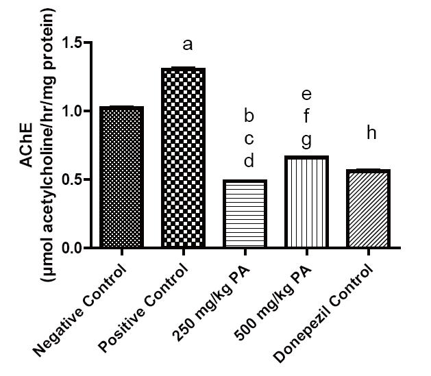
The aim of this study is to determine the ameliorative effects of methanolic extract of Platycerium angolence (MEPA)on aluminium chloride (AlCl3)-induced neurotoxicity in rats. Rats were randomly separated into five groups containing four rats each. Group 1 serves as the control group and received normal saline, group 2 rats were administered with 100 mg/kg body weight of AlCl3 orally for 28 days to induce neuronal damage, while groups 3 and 4 rats received 250 mg/kg and 500 mg/kg of MEPA for 7days after induction of neuronal damage by AlCl3 for 28 days. Group 5 rats were administered donepezil drug (0.2 mg/kg) which served as reference drug. Behavioural study such as elevated plus maze, Y-maze and open field tests were investigated on the rats. Biochemical assays such as lipid peroxidation, acetylcholinesterase activities, and metal concentration in the whole brain homogenates were estimated using standard procedures. Results revealed a significant (p<0.05) increase in the memory index (MI), lipid peroxidation of animals in the AlCl3 group when compared with the control and the MEPA treated groups. The significant (p<0.05) decrease on lipid peroxidation MEPA was dose dependent. The acetylcholinesterase activities observed in the brain of 250 mg/kg (0.49 µmol acetylcholine/hr/mg protein) group and 500 mg/kg of extract (0.66 µmol acetylcholine/hr/mg protein) has comparative effect with the group treated with the standard drug donepezil when compared with AlCl3 induced group (1.31 µmol acetylcholine/hr/mg protein). This study revealed that both doses of 250 and 500 mg/kg of MEPA has ameliorative potential against AlCl3-induced neurotoxicity in rats but 500 mg/kg of the extract shows better protection.
KEY WORDS: Neurotoxicity; Platycerium angolence; Acetylcholinesterase; Aluminium chloride|
|
INTRODUCTION |
|---|
Neurotoxicity is a form of toxicity in which a biological, chemical, or physical agent produces an adverse effect on the structure and/or functions of both the central and/or the peripheral nervous system (1, 2). Neurotoxicity occurs when there is an exposure to substances (specifically neurotoxin), which alters the normal activity of the nervous system in such a way that it causes a permanent or irreversible damage to the nervous tissue. Thus, a disruption or death of the neurons occurs. Some of the known sources of these neurotoxins are as a result of organ transplants, radiation treatment, certain drug therapies, exposure to heavy metals, pesticides, solvents, and some naturally occurring substances (3-5).
It has been well documented that food additives, therapeutic treatments and consumption of tissues of mammals such as the kidneys, liver, heart, blood, bones and brain are some of the ways humans are exposed to aluminum (6). Many aquatic invertebrates, including the brine shrimp can accumulate and tolerate a very high level of metals in their tissues. Although the concentration of metals in most saline water systems is rather low. Point source discharges such as municipal and industrial wastewater falls out which can lead to high concentrations of aluminum that affects the aquatic environment. The adverse effect of the exposure of humans to aluminum chloride (AlCl3) has been reported. AlCl3 has been documented to induce the generation of free radicals and neurotoxicity in the brain, which might lead to degenerative disorders (6-9).
AlCl3-induced neurotoxicity mouse model has been effectively used in various studies due to the fact that aluminum has high bioavailability and it can be easily administered orally and intra-peritoneally (10). Aside oxidative damage and inflammation, aluminum causes neurotoxicity by the formation of neurofibrillary tangles (11), most importantly by interacting with amyloid beta (Aβ) resulting in formation of amyloid aggregates (12).
To this end, there is the need to explore all available means to reduce the impact of neurotoxicity through natural products such as plants. Researchers have provided rich information on the application of plants in healthcare systems all over the world. However, there is paucity of scientific information on the phyto-constituents, potentials and therapeutic uses of lower plants until recently. Unlike many rainforest plant species, lower plants are not normally employed by indigenous people for healing, treatment and management of ailments (13). Platycerium angolense is one of such lower plants with acclaimed folkloric use but few scientific validations. Platycerium are epiphytes, that grows naturally on branches and trunks of trees in the tropical, subtropical and rain forests of Southeast Asia, Phillippines, Indonesia, Australia, New Guinea, Africa and South America. It is found growing in overlapping layers with serrated upper edges and stand erect forming an opened receptacle for water, dead leaves and organic debris that eventually decay and provide nutrients for the ferns (14-16). The reported pharmacological properties of Platycerium angolense includes anti-inflammatory (13), antioxidant and hepatoprotective (17, 18) activities.
However, to the best of our knowledge, no study has been carried out in the literature regarding the role of Platycerium angolense against AlCl3-induced neurotoxicity in rats. Hence, the objective of this present study, was to investigate the ameliorative effects of Platycerium angolense against neurotoxicity induced by AlCl3 in Wistar rats.
|
|
MATERIALS AND METHODS |
|---|
Plant material
The leaves of Platycerium angolense were harvested from trunks of trees around the environs of Christ Nursery School, Ado-Ekiti Metropolis, Ekiti State, Nigeria. The plant was identified by Mr. R.O. Omotayo of the Department of Plant Science and Biotechnology, Ekiti State University, Ado Ekiti, Nigeria. Specimen copy was deposited at the university herbarium and was given voucher number UHAE2018005.
Plant extraction
The plant leaves were air-dried for three weeks and pulverized into powder. Seven hundred grammes of the powdered sample was extracted in 80% methanol by maceration for 72 hours. The methanolic crude extract obtained was concentrated in a rotary evaporator, lyophilized and stored at 4oC until further use.
Experimental Animals
Adult male Wistar albino rats weighing 140 ± 5g were purchased from the animal house of the College of Medicine, Ekiti State University, Ado-Ekiti, Nigeria. The animals were acclimatized for 14 days, and given animal feed and water ad libitum. The animals were maintained under standard environmental conditions (22–25°C, 12 h/12 h light/dark cycle). All animal experiments were performed in compliance with the institutional ethics committee regulations and guidelines on animal welfare of our Institution and according to the Guide for the care and use of laboratory animals (19).
Experimental design
Twenty rats were divided into five experimental groups. Negative Control Group: received normal saline and serves as the negative control; Positive Control Group: received 100 mg AlCl3 only for 28 days to induce neurotoxicity by oral administration, 250 and 500 mg/kg groups were administered 250 and 500 mg/kg of methanolic extract of Platycerium angolense (MEPA) for seven days, after the 28 day AlCl3 induction of neurotoxicity in rats. Donepezil group were administered 0.2mg/kg standard drug (donepezil) for seven days after the 28 days of AlCl3 induction of neurotoxicity.
Induction of Neurotoxicity
Neurotoxicity was induced vial oral administration of 100 mg/kg AlCl3 for 28 days in all animal groups, except the positive control group. After the neurotoxicity induction, the animals were treated through oral administration of Platycerium angolense plant extract and standard drug. The oral administration was done via cannula during the duration of the experiment.
Behavioural Studies
Elevated Plus Maze. Elevated plus maze apparatus was designed according to the description of Adeniyi and Olatunji (20). The animal was placed in one of the closed arms in the apparatus and recording the following behaviors: total time spent in the open arm, total time spent in the closed arm, open arm entries, closed arm entries, rearing and grooming. The numbers of entries into each portion of the apparatus (open and closed) are scored in addition to the total time spent in each portion. The maze is cleaned with ethanol after every trial. Total entries score is also an index of anxiety, and the percentages of entries and time spent in each arm constitute the index of primary anxiety (21, 22).
Y-Maze. Testing occurred in a Y-shaped maze with three white, opaque wooden arms at a 120° angle from each other. After introduction of the animals to the center of the maze, the animal is allowed to freely explore the three arms. The frequencies of correct alternations (ABC, ACB, BCA, BAC, CBA, or CAB) between the arms (ABC) were recorded (manually) to determine the memory index (percentage of correct alternation) as described by Adeniyi and Olatunji (20) and Adeniyi et al. (23).
Open Field Test. The method of Walsh and Cummins (24) was used to simultaneously measure the locomotion, exploration and anxiety of the experimental animals. The open field is an arena with walls to prevent escape. The field is marked with a grid and square crossings. The center of the field is marked with a different color to differentiate from the other squares. A camcorder was placed about 150cm above the apparatus in order to capture the movement of the rats without any interference. Behavioral patterns measured in the open field test include line crossing (frequency with which the rodent crossed a grid line with all four paws), rearing (frequency with which the rodent stood on their hind legs in the field) and grooming.
Determination of Lipid peroxidation and Catalase Activities
Lipid peroxidation was assayed by measuring the formation of thiobarbituric acid reactive substances (TBARS) (expressed as MDA equivalents) described by the method of Ohkawa et al. (25). The malondialdehyde (MDA) level was calculated from the absorbance according to the method of Adam-Vizi and Seregi (26) Catalase activity Catalase (CAT) activity was measured according to the method described by Sinha (27) by means of H2O2 as substrate. The results were expressed in μmoles H2O2 consumed/ min.
Cholinergic Enzyme Activity
The acetylcholinesterase (AChE) enzymatic assay was determined using a modification of the spectrophotometric method of Ellman et al. (28) as described by Akinyemi et al. (29). The reaction medium (2 ml final volume) contained 100 mmol/l of K+-phosphate buffer, pH 7.5, and 1 mmol/l of 5,5-dithiobisnitrobenzoic acid. The enzyme (40–50 mg of protein) was pre-incubated for 2 min and the reaction was initiated by adding 0.8 mmol/l of acetylthiocholine iodide and enzyme activity expressed in µmol AcSCh per min per mg of protein.
Statistical Analysis
The data collected were analysed using one–way analysis of variance (ANOVA) followed by Duncan post hoc test. A value of p<0.05 was considered statistically significant.
|
|
RESULTS |
|---|
General Observation and Effect of AlCl3 on animal body weight
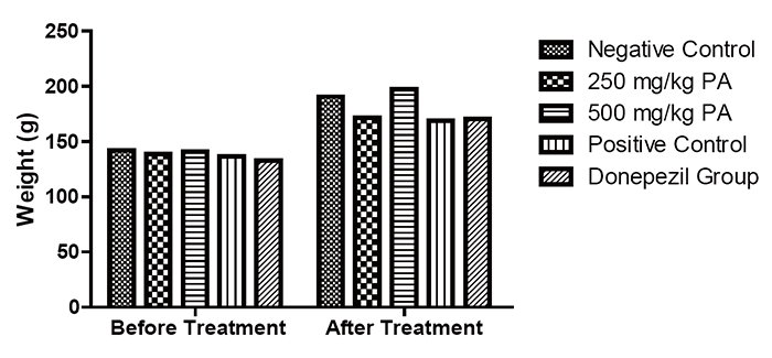
There was no observable neurological symptom (i.e. hemiplegic paralysis and seizure) and behavioural abnormalities (i.e. poor feeding, irritability and abnormal locomotor activity) seen in all groups. The Aluminum chloride treatment had no significant (P>0.05) effect on the general body weight of the experimental animals as shown in Figure 1.
Effect of treatment on behavioural parameters
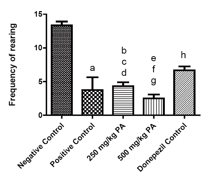
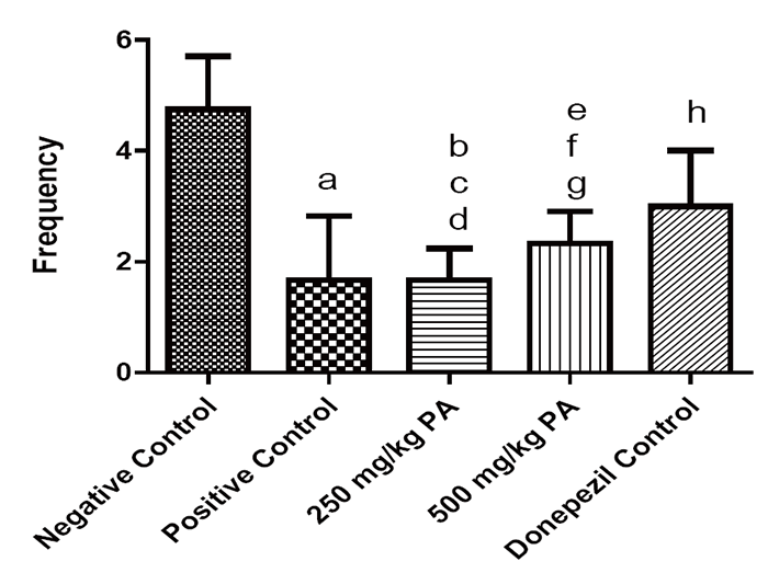
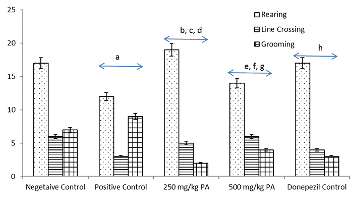
The ethological measures that were observed in the rodents in the maze were the number of rears, and head dips. From this research work, 100 mg/kg aluminum chloride significantly decreased (P<0.05) locomotors activity. This was concluded through the significant decrease in numbers of rearing and total number of head dipping compared to the positive control group (Figure 2 and Figure 3). Treatment with methanolic extract of Platycerium angolense shows no significant effect on the frequency of head dipping when compared to the positive control. The group administered with 500 mg/kg, however, has significant lower frequency of head dipping when compared to the group administered with 250 mg/kg methanolic extract of Platycerium angolense. Methanolic extract of Platycerium angolense has no statistical significant changes on the frequency of head dipping and number of rearing.
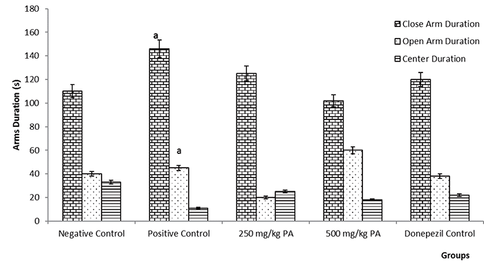
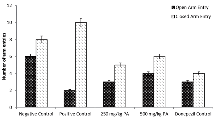
Other parameters evaluated in this test included closed and open arm duration (Figure 4) as well as open and closed arm entries (Figure 5). The positive control group has the highest level of closed arm duration and closed arm entry and by implication the lowest open arm entry, the lowest frequency of head dippings. This result implies that AlCl3 has an induced effect on brain limbic system (amygdala) that controls emotion. These features were fairly increased (but not statistically) in the open arm region of the apparatus by the Platycerium angolence methanolic extract and significantly increased by donepezil drug.
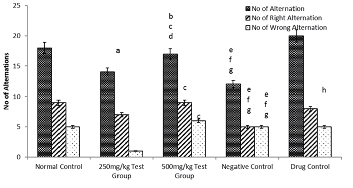
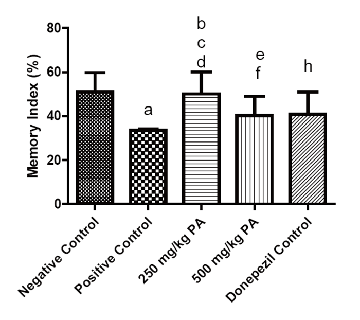
In the Y maze test, the numbers of right and wrong alternations were recorded for each group. There was significant highest total number of alternations in the group treated with donepezil drug. There was significant difference in the number of right alternation seen in the positive control group when compared to the control and test groups (groups 3 and 4). The positive control group has the lowest memory index. There is no significant difference in memory index between the negative control group and the group treated with 250 mg/kg of the plant extract. Statistically, it was noted that donepezil drug group and 500 mg/kg plant extract group showed the same effect on memory index as reflected in Figure 7.
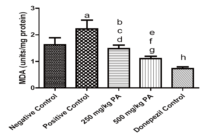
Effect on Lipid peroxidation and Catalase
The lipid peroxidation activities in the brain of the experimental animals were greatly influenced as shown in Figure 9. Lipid peroxidation was significantly (P<0.05) increased positive control when compared with the Negative Control group. When treated with the plant extract, 500 mg/kg MEPA has more reducing effect on lipid peroxidation than 250 mg/kg MEPA. The donepezil drug has significant effect on induced lipid peroxidation.
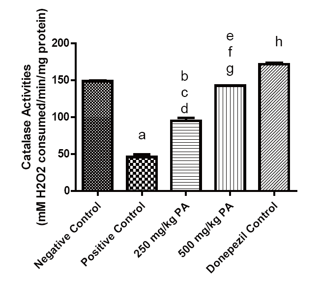
As shown in Figure 10, the catalase activity in the brain was significantly improved with the extract treatment and donepezil drug. The effect is dose-dependent.
Effect on Acetylcholime esterase activity
The acetylcholinesterase activity was evaluated and represented in Figure 11. AlCl3 induced neurotoxicity significantly increased the activities acetylcholinesterase when compared to the negative control group. The 250 mg/kg and 500 mg/kg MEPA reduced the activities of acetylcholinesterase.
|
|
|
|
|
|
|
|
|
|
|
|
|
DISCUSSION |
|---|
Aluminum chloride has been reported to cause significant short-term and long-term memory disturbances, decrease in locomotor activity, a significant inhibition of acetylcholinesterase activity in brain, significant depletion of antioxidant enzymes (catalase, glutathione reductase and glutathione peroxidase) and glutathione and significantly increased lipid peroxidation levels in cerebrum and cerebellum. Plant extract have also been employed to reduce the impact of the neurotoxicity induced by aluminum chloride. Extracts of different concentration have been reported to be a potential formulation which can be used for the treatment of Al neurotoxicity and efficient for the recovery from the toxicant-induced oxidative damage, histopathological changes and AChE activity inhibition (30, 31).
The elevated plus maze is a widely used behavioural assay for rodent and it has been validated to assess the anti-anxiety effect of pharmacological agents and steroid hormones, and to define brain regions and mechanism underlying anxiety-related behavior (32).
Adeniyi and Olatunji (20) reported that the open field test provides simultaneous measures of locomotion, exploration and anxiety. The number of line crosses and the frequency of rearing are usually used as measures of locomotor activity, but also measures of exploration and anxiety. A high frequency of these behaviours indicates increased locomotion and exploration and/or a lower level of anxiety. The number of central square entries and the duration of time spent in the central square are measures of exploratory behaviour and anxiety. A high frequency/duration of these behaviours indicates high exploratory behaviour and low anxiety levels (20). The results from this present work revealed that AlCl3 significantly (P<0.05) reduced the spontaneous locomotor activity in the positive control group after daily treatment with 100 mg/ml AlCl3 by reducing the total number of rearing and line crossing recorded from the experimental animals (Figure 8). However, all the groups treated with the various concentrations of methanolic extract of Playcerium angolence (250 and 500 mg/kg) showed an increase in the measured parameters. This suggests that Playcerium angolence may improve locomotion and exploration activities under induced neurotoxicity condition in the rats. In a research carried out by Asuquo et al (33) on the effect of ethanolic extract of Spondias mombin on locomotor activity, it was deduced that it has a decreased locomotor activity on the experimental rats which is an indicative of depressant activity on the central nervous system (33). This suggest that MEPA cannot serve as a depressant but a stimulant.
The significant role of free radical mediated oxidative damage in the pathogenesis of neurodegenerative disorders has been firmly established (34). In particular, markers of lipid peroxidation have been found to be elevated in brain tissues and body fluids in several neurodegenerative diseases, including Alzheimer disease (AD), Parkinson disease (PD), amyotrophic lateral sclerosis (ALS), Huntington disease (HD) and Down syndrome (DS) (35). Consonant with these findings, several reports have documented increased levels of reactive products of lipid peroxidation in diseased regions of brain, but generally not in regions uninvolved in the disease (34, 35).
Lipid peroxidation is one of the major sources of free radical-mediated injury that directly damages neuronal membranes and yields a number of products responsible for extensive cellular damage (36). As shown in Figure 9 from this study, there is a significant increase in MDA levels in the brain of the AlCl3 (positive control) group when compared with their corresponding normal control group. However, the level of MDA in the extract treated groups (250 and 500mg/kg) was significantly lowered when compared to the AlCl3 only group. Inference from this is that AlCl3 induced oxidative stress in the brain of the experimental rats. This lowering effect of MDA contents is comparable to that of the reference group that was treated with donepezil. The ability of the methanolic extract of. Platycerium angolense to inhibit the observed lipid peroxidation in the brain homogenate is likely due to the abundance of flavonoids and phenolic compounds (rutin, quercetin, gallic acid) present in them as previously reported by (37). These phenolics which are secondary metabolites in plants and have been associated to several health benefits in plants which includes antioxidant, antiglycemic, anticarcinogenic, anti-inflammatory and vasodilatory properties (38-42). Furthermore, it has been reported that extract of P. angolense contains potentially useful phytochemicals and exhibits remarkable therapeutic activities. These phytochemicals could contribute to the overall antioxidant potential observed in the extract (13, 18). The ability of Platycerium angolense extracts to be able to improve catalase activities in the brain of neurotoxicity induced rats (as shown in Fig. 10) may be as a result its constituent antioxidants properties as evidenced by the presence of its phenolics (18). Noteworthy from this experiment, is that at 500 mg/kg dose, the plant extract has more significant catalase activities when compared to 250 mg/kg dose and the other experimental groups.
Acetylcholinesterase is a remarkably efficient serine hydrolase responsible responsible for the termination of impulse signaling at cholinergic synapses (43). Its principal biological role is termination of impulse transmission at cholinergic synapses by rapid hydrolysis of the neurotransmitter acetylcholine to acetate and choline. In certain neurological disorders such as Alzheimer’s disease (AD), acetylcholinesterase is over activated in the synapses so that levels of acetylcholine in the brains is significantly diminished, which leads to weakened neurotransmission and thereby memory loss and other adverse effects (43-45). The cholinesterase enzyme may also be, at least partly, responsible for the buildup of amyloid β plaques and the neurofibrillary tangles in AD brains. This study shows significant increased activities of the acetylcholinesterase enzyme in the brain of the positive control group when compared to the negative control (Figure 11). However, the lowered acetylcholinesterase enzyme activiteis observed in the extract treated groups maybe as a result of the ability of Platycerium angolense to scavenge or inhibit acetylcholinesterase enzyme. It was suggested that anti-acetylcholinesterase properties of L. officinale and B. integrima may offer great potentials for the treatment of different neurological diseases including AD (46). In this manner, P. angolense may therefore be suggested as alternative therapy for the treatment and/or management of diseases induced by neurotoxicity.
|
|
CONCLUSION |
|---|
In this study, AlCl3 induced neurotoxicity at 100 mg/kg body weight of albino rats. The methanolic extract of P. angolense showed no significant effect in the parameters (head dips and rearing) measured using elevated plus maze but may improve locomotion and explorative activities as measured with the open filed test apparatus. The plant extract showed significant antioxidant ability in reducing lipid peroxidation (MDA level) and increasing catalase activities in the brain. The extract also had an inhibitory effect on the activities of acetylcholinesterase. Thus, it suggests that methanolic extract of Platecrium angolence has ameliorative potentials against induced neurotoxicity in rats. However, much work is still required on the isolation of the active components of the plant for further in vivo studies on the different compartment (hippocampus, cerebral cortex, and cerebellum) of the brain.
|
|
CONFLICT OF INTEREST |
|---|
The authors declare that no conflicting interests exist.
|
|
FUNDING SOURCES |
|---|
This research did not receive any specific grant from funding agencies in the public, commercial, or not-for-profit sectors.
|
|
REFERENCES |
|---|