| ORIGINAL ARTICLE |
|
|
Department of Prosthodontics, M R Ambedkar Dental College, Bangalore, India
Corresponding Author: Dr. Vinaya Kundapur, Reader, Department of Prosthodontics, M R Ambedkar Dental College, Bangalore, India. Mobile: +91 8904073488; E-mail: drvinni@gmail.com.
| |
ABSTRACT |
| INTRODUCTION | |
|
|
REVIEW OF LITERATURE |
|
|
MATERIALS & METHODS |
|
|
METHODOLOGY |
|
|
RESULTS |
|
|
CONCLUSION |
|
|
REFERENCES |
|
|
ABSTRACT
|
|---|
Background and Objectives: Midline fracture of dentures and fractures due to sudden impact on denture bases, like accidental dropping, are common. The limitation that the strength of these materials displays sets a need for further improvement of the polymethylmethacrylate resin. Studies have demonstrated that carbon nanotubes possess outstanding mechanical properties of flexural and impact strengths. The present study evaluated and compared the flexural strength and surface roughness on reinforcing conventional heat cure denture base resins and high impact resins with carbon nanotubes. Method: A metal template of specific dimensions 65 mm × 10 mm × 3 mm was fabricated and flasked to obtain moulds. Monomer (methylmethacrylate) containing 1wt% carbon nanotubes was mixed with DPI and Lucitone polymers respectively and packed into the moulds to obtain 20 samples for each. Monomer without nanoparticles was used to fabricate 20 more samples for each resin. Following curing and deflasking, a total of 80 specimens were finished to required dimensions. Surface roughness of each specimen was evaluated using an optical profilometer. The flexural strengths were evaluated using a Universal Testing Machine. Results: The results showed that there was statistically significant difference among the flexural strength values of the groups with microadditions of CNTs when compared to the groups without microadditions. The surface roughness values, on comparison, showed no statistical significance. Interpretation and Conclusion: On comparison of the values obtained, the study concludes that flexural strength significantly improved on addition of carbon nanotubes. However, the comparison of surface roughness values suggested that incorporation of nanotubes does not increase the surface roughness of denture resins.
KEY WORDS: polymethylmethacrylate; reinforced resins; carbon nanotubes; nanoparticles; flexural strength; surface roughness|
|
INTRODUCTION |
|---|
Polymethylmethacrylate has long since been the resin of choice for fabrication of denture bases in clinical dentistry because of its favorable working characteristics, processing ease, accurate fit, stability in the oral environment and superior esthetics (1). Despite these excellent properties, midline fracture of dentures and fractures due to sudden impact on denture bases, like accidental dropping, are common. The midline fracture in dentures is often the result of flexural fatigue (2). The limitation that the strength of these materials displays sets a need for further improvement of the polymethylmethacrylate resin.
The reinforcement of denture base material has been a subject of interest to the dental material community (3). Various materials have been evaluated as reinforcers in Polymethylmethacrylate to enhance the mechanical properties. Nanotechnology has the potential to bring enormous changes into the fields of medicine and dentistry (4). Carbon nanotubes are strong, resilient, and lightweight, and usually form stable cylindrical structures (3). Nanomaterials like carbon nanotubes are allotropes of carbon with a cylindrical structure. Studies have demonstrated that carbon nanotubes possess outstanding mechanical properties of flexural and impact strengths.
The purpose of this study is to evaluate and compare the mechanical properties of flexural strength and surface roughness on reinforcing nanomaterials like carbon nanotubes in conventional heat cure resins and high impact denture base resins.
|
|
REVIEW OF LITERATURE |
|---|
A study was conducted to compare the flexural and impact strengths of 3 commercially available light activated denture base resins- Eclipse, Triad Visible light cure and Lightplast, after reinforcing them with single walled carbon nanotubes. The flexural strength of each material also increases gradually with increasing percentage of addition of single walled carbon nanotubes. The results demonstrated that Eclipse denture base showed better impact and flexural strengths than other light cured reinforced denture base acrylic resins. Also the flexural strength of each material increased with increasing percentage of addition of single walled carbon nanotubes (5).
A study to compare the polymerization shrinkage of denture base acrylic resin with and without microadditions of carbon nanotubes was carried out. Two materials were used, polymethylmethacrylate resin and multiwalled carbon nanotubes. Four groups were established of 10 specimens each according to the weight percent of carbon nanotubes dispersed and disintegrated in the monomer: group I (0.5% of carbon nanotubes in monomer), II (0.25%), III (0.125%), and IV (control group, 0%). The polymerization shrinkage of acrylic resin for each group was evaluated based on the distance between the reference points in wax (before polymerization) and in acrylic (after polymerization), measured using a traveling microscope. The data were submitted to Kruskal-Wallis and one-way ANOVA for comparison among the groups, and the results were statistically analyzed. Results obtained showed that the order of severity of polymerization shrinkage was 0% > 0.125% > 0.25% > 0.5% for the amount of carbon nanotubes incorporated in methylmethacrylate (6).
A study evaluated the effect of incorporating carbon nanotubes and graphene on polymerization shrinkage of heat cure acrylic resin. Standard edentulous maxillary casts were made to fabricate different types of denture bases. On the cast, wax templates in the shape of posterior palatal seal were made to standardize the quantity of modified material incorporated in the posterior palatal seal area i.e. acrylic mixed with different concentrations of carbon nanotubes or graphene. After denture base fabrication, denture base and cast was trimmed together in the posterior region and the gap between cast and denture base was measured using a stereomicroscope. The gap between cast and denture base was found to be least with the 0.5% carbon nanotube group (0.09 mm). Thus the study concluded that when the concentration was increased polymerization shrinkage was observed to decrease. Addition of carbon nanotubes and graphene in denture base acrylic resin showed reduction in polymerization shrinkage (7). A study was conducted to evaluate the transverse strength, impact strength, surface hardness and surface roughness test of high impact heat cure resin with 1 wt % of carbon nanotubes. This was preceded by a pilot study using different concentrations of carbon nanotubes- 0.5%, 1%, 1.5% in which 1 wt% was found to be the best concentration and thus was used in the study. 10 ml of monomer is added to 21 mg of powder after mixing, and reaching the dough stage, applying it carefully into the mould, which were previously fabricated in the lab following the conventional flasking technique for complete denture. There was a significant increase in impact strength and transverse strength when carbon nanotubes in 1wt% were added, compared to control group where as surface hardness decreased when adding carbon nanotubes (8).
|
|
MATERIALS & METHODS |
|---|
A total number of 80 samples will be divided into 4 groups:
Grouping of specimens will be done as follows:
Group I: Conventional heat cure polymethylmethacrylate with no microadditions (control group I);
Group II: High impact polymethylmethacrylate with no microadditions (control group II);
Group III: Conventional heat cure polymethylmethacrylate with carbon nanotubes 1wt%;
Group IV: High impact polymethylmethacrylate with carbon nanotubes 1wt%.
|
|
METHODOLOGY |
|---|
A template of specific dimensions 65 mm × 10 mm × 3 mm for fabrication of wax blocks of standard size will be fabricated. Wax blocks will be fabricated using standard pink modelling wax and flasking done followed by dewaxing to create molds for the heat cure resins. Electronic precision balance will be used to weigh multiwalled carbon nanotubes 1wt % respectively.
Carbon nanotubes would be subjected to ultrasonic agitation in an ultrasonic unit for 5 minutes for uniform dispersion in the monomer (methylmethacrylate), taking care not let the particles agglomerate. Monomer containing carbon nanotubes fillers will be mixed with polymer of each resin respectively in standard 1:3 ratio and flasked. Monomer with no microadditions will act as control group. Curing will be done by heating the flask at 65°C for 90minutes, then at 100°C for 1 hour for the conventional heat cure and high impact resins. Bench cooling will be done for 3 hours after which specimens will be deflasked. The specimens will be finished to the required dimensions. Each specimen will then be mounted on a jig secured firmly at its ends and a three point flexural test will be done using Instron universal testing machine. Surface roughness will be evaluated using a profilometer.
The values of flexural strength will be obtained using the Universal Testing Machine and a profilometer will be used to estimate the surface roughness for each specimen. Statistical analysis of collected data will be done.
By using the Universal Testing Machine, flexural strength will be evaluated and a profilometer will be used to estimate the surface roughness for each specimen. Data will be analyzed statistically with one way ANOVA and post hoc Tukey analysis using statistical software of a personal computer.
|
|
RESULTS |
|---|
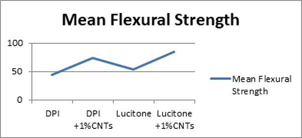
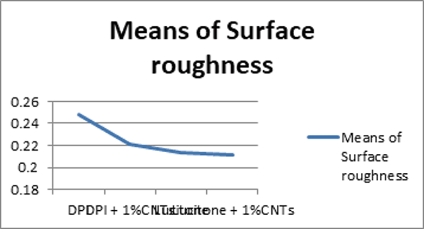
The present study was done to compare the flexural strength and surface roughness of 4 groups of denture bases:
Group 1: DPI;
Group 2: DPI+ 1wt% MWCNT;
Group 3: LUCITONE;
Group 4: LUCITONE +1wt% MWCNT.
From the ANOVA table it is noticed that the values for flexural strength of the four groups of denture bases denote a highly significant difference (P<0.001) among all the four groups. (p<0.001).The difference exhibited by surface roughness of all four groups on the other hand is found to be insignificant after ANOVA analysis (p>0.001). In order to find out among which pair of groups of denture bases there exists a significant difference with respect to flexural strength, a post hoc Tukey analysis was done.
The post hoc Tukey analysis showed a significant difference between all the four groups after pairwise comparison for flexural strength. It was found that DPI exhibited lowest values in terms of flexural strength. Lucitone performed better than DPI in terms of flexural strength. Lucitone denture base material with 1wt% microadditions of carbon nanotubes was found to be the best amongst the four groups, in terms of flexural strength, followed by DPI with microadditions of 1wt% carbon nanotubes.
As ANOVA analysis was found to be insignificant for surface roughness comparison between the four groups, a post hoc test was not required and would be unnecessary as results from the one- way ANOVA were conclusive enough.
|
|
CONCLUSION |
|---|
Within the limitations of this study, the following conclusions were drawn:
1. Incorporation of 1wt% CNTs into PMMA resin produced a statistically significant increase in flexural strength values. Both the commercially available polymethylmethacrylate resins when reinforced with nanotubes performed better when subjected to the UTM, as compared to their control groups. Lucitone with carbon nanotubes were found to be the best followed by DPI with nanofillers. DPI denture base resins without incorporations produced the least values for flexural strength, followed by Lucitone without any nanofillers (Table 1) (Figure 1).
2. On comparison of values obtained for the groups for surface roughness, a slight decrease in the surface roughness values was seen after addition of nanotubes. However, this finding is not statistically significant. Hence carbon nanotubes do not affect the surface profile of resins, thus being a suitable filler of choice to reinforce polymethylmethacrylate denture base material (Table 2) (Figure 2).
The continuation of this research is of utmost important due to the promising nature of carbon nanotubes as a strengthening agent to reinforce PMMA. Further future studies would open the opportunity to explore a vast range of utilities of this miraculous strengthener.
|
|
|
|
|
|
|
REFERENCES |
|---|