| ORIGINAL ARTICLE |
|
|
1 Department of Dental Public Health, Dhaka dental college, Dhaka, Bangladesh;
2 Nutrition and Clinical Service Division, International Centre for Diarrheal Disease Research, Bangladesh (ICDDR'B), Mohakhali, Dhaka-1212, Bangladesh;
3 Medical officer, DGHS, Dhaka, Bangladesh;
4 Department of Microbiology, Jahangirnagar University, Savar, Bangladesh;
5 Dhaka Medical College, Dhaka, Bangladesh;
6 National Institute of Preventive and Social Medicine (NIPSOM), Mohakhali, Dhaka-1212, Bangladesh
Corresponding Author: Md Haroon Or Rashid, Nutrition and Clinical Service Division, International Centre for Diarrheal Disease Research, Bangladesh (ICDDR’B), Shaheed Tajuddin Ahmed Sarani, Mohakhali, Dhaka-1212, Bangladesh. E-mail: haroon9330@gmail.com.
| |
ABSTRACT |
| INTRODUCTION | |
|
|
METHOD |
|
|
RESULT |
|
|
DISCUSSION |
|
|
CONCLUSION |
|
|
CONSENT |
|
|
ETHICAL APPROVAL |
|
|
CONFLICT OF INTEREST |
|
|
REFERENCES |
|
|
ABSTRACT
|
|---|
Introduction: In 1941 Sodium fluorite containing tubes was developed. Since then these tables were used in the clinical laboratory for measurement of glucose. Sodium fluoride is a useful agent that inhibits glycolysis. The glucose level in the unpreserved blood sample can decrease by 5-7% per hour due to glycolysis. Methods: The objective to evaluate the stability of blood glucose in fluorinated plasma and plane tube serum. The Blood samples obtained from 30 participants and which pour into two different (fluorinated and plane) tube. Plasma and serum separated and storage within 25-30 Minute of the collection on average. Separated samples were aliquoted into eppendorfs and stored in the different cool box at 4-8oC. Glucose concentrations were analyzed by glucose oxidase method using the SELECTRA Pro-M Auto-analyzer at 0hours (within 30 min), 24hours and 48 hours. Paired sample t-test performed significantly at a P-value of less than 0.05. Results: Results show that the rate at which glucose concentration changed with time varies in fluorinated plasma and plane tube serum, the average mean difference of glucose 1.2 mg/dl, 9.2 mg/dl, and 3.32 mg/dl, 3.11 mg/dl in plasma and serum from 0 h-24 h and 0 h-48 h respectively. A statistically significant correlation (p<0.05) found with times of measurement. Glucose levels slightly reduce in plasma than serum within 24 h but comparatively more glycolysis occurs in plasma than serum after 24 hour. Conclusion: This study tries to find out a suitable agent for assessing blood glucose levels when the analysis delayed. Sodium fluoride is an effective agent for delay glucose analysis. .
KEY WORDS: Blood Glucose; Sodium fluorinated tube; Plane tube; Duration|
|
INTRODUCTION |
|---|
Glucose obtained from Metabolism of carbohydrate, a certain amount of glucose present in a mammals blood. The normal blood range of rage of glucose 4-6 mmol/L (1). The glucose concentration in unpreserved blood samples decreases rapidly 5%–7% per hour due to glycolysis (2). A suitable anticoagulant prevents coagulation of blood and reduces the loss of glucose (3). In 1941, Sodium fluoride containing tubes introduced, for measuring glucose in the clinical laboratory. Sodium fluoride (NaF) has an anti-glycolytic effect that inhibits glycolysis by erythrocytes (4). However, where the blood samples collected in the field site and several hours may require reaching the laboratory for laboratory analysis. NaF is not an effective agent to prevent glycolysis. Sodium fluoride (NaF) inhibits enolase, an enzyme in the glycolytic pathway. Although NaF has been shown to completely arrest glycolysis by four hours, it has little effect on the rate of glycolysis during the first 1-2 hours (5). It recognized that the blood sample subjected to in vitro glycolysis, which enhanced by the presence of leucocytes and diminished by sample refrigeration (6). Many researchers have concluded that the addition of sodium fluoride to blood sample is capable, in at least some parts of decreasing ex vivo glycolysis which results to decline in glucose concentration of the blood sample (7). Fluoride oxalate is an anticoagulant which binds to ionized calcium, thereby preventing blood from clotting (8). Sensitive and stable analytical methods for measurement of serum glucose levels developed which include the hexokinase-glucose-6-phosphate dehydrogenase and glucose oxidase assays (9).
Recently, Sanders and Deadman using a fairly precise glucose analyzer, reported that NaF was able to preserve glucose in whole blood for 48 h. Their findings were promptly disputed (10). In view of the controversy and the inevitable delay in the separation of plasma, we have studied in greater detail the changes in glucose concentration with time in blood samples with and without added NaF (11).
|
|
METHOD |
|---|
This study was performed originally as part of quality assurance of project to determine what blood tubes are best suited to give accurate and stable serum/plasma glucose levels, i.e., which tube type allowed maximum reproducibility upon storage. A cross-sectional study was conducted at the SAIC DIGITAL Laboratory from 20 May – 22 May 2018 (72 hours). Random and fasting blood sample was collected from 30 voluntary participants. A volume of 2 ml and 3 ml whole blood was collected in potassium oxalate tube containing Sodium Fluoride-NaF (for plasma) and plain tube (for serum) respectively. Collection time was 8:30 Am to 12:20 PM. Plasma and serum were separated within 40-50 minute of collection on average. Separated plasma and serum were aliquoted into three sets of eppendorfs labeled as 0.0 hours, 24 hours and 48 hours separately. Following, labeled plasma and serum were stored in two different cold boxes labeled as 24 hours and 48 hours with ice pack along and a dial thermometer for temperature monitoring. Glucose concentrations were measured by glucose oxidase assays method using the SELECTRA Pro-M, Auto-analyzer at 0hours (within 30 minutes after aliquote), 24 hours and 48 hours. Results were enumerated in the datasheet and also inputted into SPSS software for statistical analysis.
|
|
RESULT |
|---|
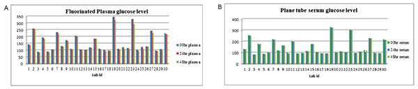
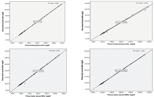
Comparing glucose level in fluorinated plasma and plane tube serum, results of glucose values (n=30, paired t-test) showed that there was a significant difference. The mean ± standard deviations (SD) of glucose concentrations were 153.07 ± 70.57 mg/dl, 151.95 ± 70.56 mg/dl, and 143.86 ± 65.48 mg/dl in fluorinated plasma and coefficient of variance were 46.10%, 46.43% and 45.51% at 0hour, 24 h, and 48 h respectively. Average deference after 0 h to 24 h was 1.2 mg/dl and p-value 0.001 and average deference after 0h to 48h was 9.2 mg/dl and p-value 0.001. On the contrary, the mean ± standard deviations (SD) of glucose concentrations were 143.76 ± 66.97 mg/dl, 140.44 ± 66.48 mg/dl and 140.65 ± 66.40 in plain tube serum and coefficient of variance were 46.58%, 47.33% and 47.20% at 0 hour, 24 h, and 48 h respectively. Average deference after 0 h to 24 h of 3.32 mg/dl and p-value 0.001 and average deference after 0 h to 24 h of 3.11 mg/dl and p-value 0.001. The statistic shows that mean deference of blood glucose level were 9.30 mg/dl in fluorinated plasma and plane tube serum at 0 hour (Table 1) (Figure 1 and Figure 2).
|
|
|
|
|
DISCUSSION |
|---|
This paper highlights the number of glucose levels reduced with or without preservation of whole blood at 0 h to 24 h and 0 h to 48 h. The aim of this study was to the stability of blood glucose in fluorinated plasma and plane tube serum. This study found that statistically significant difference in glucose level at 0 h, 24 h and 48 h in measurement. The mean difference was 1.2 mg/dL at 0 h to 24 h and 9.20 mg/dL at 0 h to 48 h in the fluorinated tube and P-value 0.001 which obtained from paired sample t-test (12). On the contrary, the mean difference of serum glucose was 3.32 mg/dL and 3.11 mg/dL at 0h to 24 h and 0 h to 48 h respectively. These results are comparable with (13) they have found the mean difference range from 6.1 mg/ dL to 7.4 mg/dL though we cannot truly compare our results with their study as the composition of commercially available citrate buffer tubes used by them may be different from the tubes prepared by us. This study also found that glucose level relatively slowly reduces after 24 hours.
In this study, we found that the lowest CVs for fluorinated plasma glucose concentration was 46.10% at 0 h and CVs was 46.58% in-plane tube serum at 0 h. The result is consistent with Gupta S et al 2016, where height CVs was 14.0% in gray tubes (14).
Finally, for the measurement of serum or plasma glucose by use of plane tubes in preference to fluorinated tubes. So these tubes can be used universe for the majority of biochemistry and immunology tests.
|
|
CONCLUSION |
|---|
We conclude that there was a difference in glucose values in fluorinated plasma and plane tube serum, several times. Sodium fluoride is an effective agent for preserving glucose, having a slight effect in the first 0 h to 24 h but slowly glycolysis occur after 24 h but the contradictory result found in serum, glucose level relatively slow reduce after 24 h. However, fluorinated plasma is more effective than plane tube serum for analysis of blood glucose within 24 hours of collection.
|
|
CONSENT |
|---|
As per university standard guideline participants’ consent have been collected and preserved by the authors.
|
|
ETHICAL APPROVAL |
|---|
As per university standard guideline ethical approval has been collected and preserved by the authors.
|
|
CONFLICT OF INTEREST |
|---|
The authors declare that no conflicting interests exist.
|
|
REFERENCES |
|---|