| CASE REPORT |
|
|
1 Private Dental Practice, Cagliari, Italy;
2 Freelancer in Piacenza, Italy;
3 University of Milano, Departement of Oral Radiology, Milano, Italy
Corresponding Author: Cinzia Casu, Private Dental Practice, Cagliari, Italy. E-mail: ginzia.85@hotmail.it.
| |
ABSTRACT |
| INTRODUCTION | |
|
|
CASE REPORT |
|
|
DISCUSSION |
|
|
CONFLICT OF INTEREST |
|
|
REFERENCES |
|
|
ABSTRACT
|
|---|
Aim: To report a clinical presentation, diagnosis and treatment of a particular case of traumatic fibroma associated an occlusal defect. Background: Fibroma is benign neoplasm, whose causative agent is represented by a chronic or a traumatic stimulus. Case report: A 52-year-old healthy woman, came to our observation with a condition of an increased volume of the gingival tissue in the region between the back of the upper central incisors and the palatine wrinkles. In the middle of the exophytic lesion, could be noted invaginations that corresponded to the incisal edges of the lower incisors. An orthopantomoghraph and Dental Scan was performed that showed a reduction of the bone tissue and the thinning of the corresponding gingival cortex, to the area of incisal traumatism. The hystological examination confirmed the presence of an inflammatory hyperplasia, with traumatic etiology. Conclusion: The patient was advised to the use of a soft resin bite to reduce occlusal trauma and was sent to a gnathologist for an appropriate treatment plan.
KEY WORDS: Traumatic fibroma; Focal fibrous hyperplasia; pyogenic granuloma; peripheral giant-cell granuloma; peripheral ossifying fibroma; mucocele|
|
INTRODUCTION |
|---|
The oral tissues are exposed to continuous traumatic and phlogistic insults, the most affected areas are the tongue, palate, vestibular mucosa and periodontal tissues (1). Reactive lesions are tumor-like hyperplasia which show a response to a low-grade irritation or injury, such as chewing food impaction, calculus, and iatrogenic injuries (broken teeth, overhanging dental restorations and extended flanges of denture) (2, 3).
The reactive lesions of the oral cavity are: irritation fibroma, pyogenic granuloma, peripheral giant cell granuloma and cemento-ossifying fibroma. Other reactive lesions are epulis fissuratum inflammatory papillary hyperplasia and inflammatory fibrous hyperplasia (1).
In a retrospective study of Hamideh K. et al, the lesions were classified into two groups: fibrous lesions with connective tissue predominantly consisting of collagen (irritation fibroma, giant cell fibroma, epulis fissuratum, peripheral ossifying fibroma) and soft hemorrhagic lesions that are highly vascular (pyogenic granuloma, epulis granulomatosum, peripheral giant cell granuloma and pregnancy tumor). In this study the prevalence of reactive lesions was 20.2% and the most common peripheral lesion was pyogenic granuloma (4).
Most studies have found an increased presence of these lesions in women than men. This difference may be due to the role of hormonal factors as predisponding factors in the development of these lesions (5, 6).
The traumatic fibroma, also know as a focal fibrous hyperplasia (FFH), is considered the most common benign tumor in the oral cavity (7), it consists in a reactive hyperplasia of connective tissue. It presents a superficial or deep location and there are different types of fibroma, depending on its origin that can be odontogenic, not odontogenic (8). It is observed with a higher frequency between 40 and 60 years of age. It presents itself clinically as a nodule of variable dimensions and of rosy or white color, as result of hyperkeratosis due to traumatism (1).
Fibroma can be sessile or pedunculated. The most common site is the vestibular mucosa, tongue, gingiva and palate, associated with the reaction of chronic trauma, such as chewing on the cheeks, cheilophagia, amalgam fractured or irritation by prothesis (9). In many cases fibroma has to do with defective acrylic overlays or misfitting dentures that irritate the palate, inducing a pathological overgrowth of the fibroblasts and the collagen produced by them, which causes a submucosa mass evident on clinical examination (10). In most cases the lesion is asymptomatic and the size can vary from a few millimeters to several centimeters in diameter. Treatment of the fibroma involves surgical excision using scalpel and laser, recurrences are very infrequent (1).
Definitive diagnosis is based on histological analysis to rule out the possibility of lesions that may have a similar appearance, such as, pyogenic granuloma (PG), peripheral giant- cell granuloma (PGCG) and peripheral ossifying fibroma (POF) and mucocele (11, 12, 13, 14).
|
|
CASE REPORT |
|---|
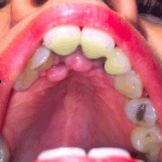
A 52-year-old woman patient came to our observation for a dental check-up. The anamnesis reported a good general state of health, while at the oral level emerged previous fixed prosthetic rehabilitations on the upper central incisors, outcomes of conservative and endodontic therapies and the lack of some dental elements. Furthermore, the patient presented a malocclusion with an increased overjet and overbite. Observing the oral mucosa it was found the presence of an increase in volume of the gingival tissue in the region between the back of the upper central incisors and the palatine wrinkles (Fig. 1).
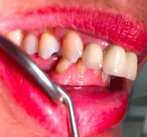
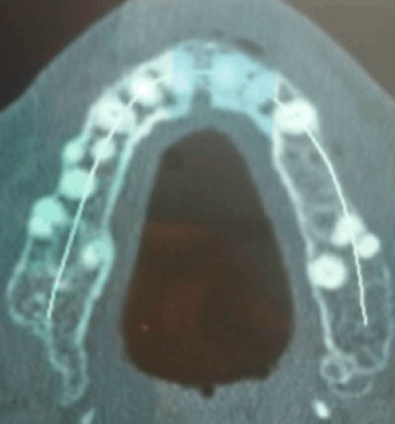
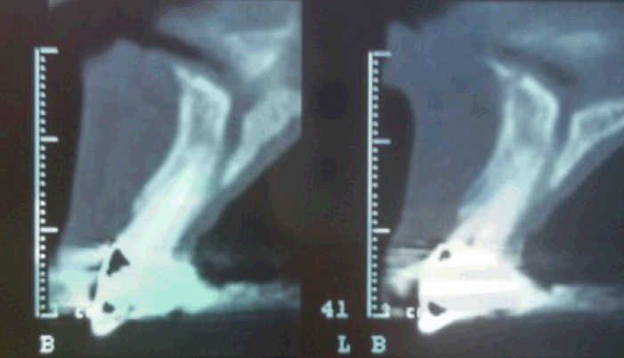
Furthermore, in the middle of the exophytic lesion, could be noted invaginations that corresponded to the incisal edges of the lower incisors. Going to reevaluate the patient's occlusion, the lower incisors in occlusion went to traumatize the front of the palate (Fig. 2). The dental orthopantomography didn’t show any type of lesion, therefore a Dental Scan was performed to verify the presence of a bone lesion (Fig. 3-4).
The three-dimensional examination showed a reduction of the bone tissue and the thinning of the corresponding gingival cortex, to the area of incisal traumatism.
It was decided to subject the patient to an incisional biopsy for a precise diagnosis. Histological examination highlighted the presence of an inflammatory hyperplasia, with possible traumatic etiology. The patient was advised to use a soft resin bite to reduce occlusal trauma and was sent to a gnathologist for an appropriate treatment plan.
|
|
|
|
|
|
DISCUSSION |
|---|
The term “inflammatory hyperplasia” is used to describe a large range of commonly occurring nodular growths of the oral mucosa that histologically represent inflamed fibrous and granulation tissue (11).
Reactive hyperplastic lesions are relatively common in centers of oral pathology (4). In according to the study of Kadeh et al., the prevalence of reactive lesions are 20.0% and the most common lesions are pyogenic granuloma and irritation fibroma. These lesions are more frequent in women (60%) than men (40%) and the most common locations of involvement are the gingiva and alveolar mucosa of the mandible (4).
The fibroma is a benign mesenchymal neoplasm that appears very frequently in the cavity. Within these lesions, a group of reactive hyperplasia that develop in response to a chronic, recurring tissue injury stimulates an exuberant or excessive tissue repair response (15).
Most fibromas represent reactive focal fibrous hyperplasia due to trauma or local irritation, although this term is more accurately describes the clinical appearance and pathogenesis of this entity, it is not commonly used.
By histological examination it was possible to find the presence of a traumatic fibroma, marked by the presence of the stratified squamous epithelium of variable thickness, below which there may be dense fibrous connective tissue with abundant collagen fibers, interspersed with fibroblasts, fibrocytes and small vascular spaces.
The fibroma is the most common non-neoplastic growth in the oral cavity. It has been know as irritation fibroma, fibrous hyperplasia, traumatic fibroma, focal fibrous hyperplasia, localized hyperplasia, fibrous polyp (16) and fibroepithelial polyp. The epidemiology of most non-neoplastic growths in the oral cavity are similar and the identification depends on histopathological differentiation.
The differential diagnoses will depend on the size and location of the lesion and they can be: papilloma, lipoma, neurofibromas, salivary gland tumor, giant cell peripheral granulomas. If they reach to develop giant sizes, their distinction will be with large mucoceles, peripheral odontogenic fibromas and spinocellular carcinomas (17).
Although conservative surgical excision and removal of causative irritations (plaque, calculus, foreign materials, source of trauma) are the usual treatments for gingival lesions, the excision should extend down to the periosteum and the adjacent teeth should be thoroughly scaled to remove the source of continuing irritation (11).
Excisional surgery is the treatment of choice for gingival hyperplasia, but some new approches for treatment such as cryosurgery, excision by Nd:YAG laser, flash lamp pulsed dye laser, injection of ethanol or corticosteroid and sodium tetradecyl sulfate sclerotherapy have been reported as alternative therapies (11).
Focal fibrous hyperplasia is a slowly progressing lesion, the growth of which is generally limited, so due to the lack of symptoms, patients will be treated long after the injury has started.
It is important to perform a careful differential diagnosis and a long-term follow-up of the case is necessary to prevent any relapse.
|
|
CONFLICT OF INTEREST |
|---|
The authors declare that no conflicting interests exist.
|
|
REFERENCES |
|---|