| ORIGINAL ARTICLE |
|
|
1 ENT Department, Ekiti State University Teaching Hospital, Ado Ekiti, Nigeria;
2 ENT Department, Federal Teaching Hospital Ido-Ekiti/Afe Babalola University, Ado-Ekiti, Nigeria;
3 Radiology department, Ekiti State University Teaching Hospital, Ado-Ekiti, Nigeria;
4 Department of Anatomic Pathology, Ekiti State University Teaching Hospital, Ado-Ekiti, Nigeria
Corresponding Author: Dr. Shuaib Kayode Aremu, ENT Department, Federal Teaching Hospital Ido-Ekiti/Afe Babalola University, Ado-Ekiti, Nigeria. Phone: +2348033583842; E-mail: shuaib.aremu@gmail.com.
Running title: CLINICOEPIDEMIOLOGICAL PRESENTATION OF SINONASAL MASS IN A NIGERIAN TERTIARY HEALTH CARE CENTRE
| |
ABSTRACT |
| INTRODUCTION | |
|
|
MATERIALS AND METHODS |
|
|
RESULTS |
|
|
DISCUSSION |
|
|
LIMITATIONS |
|
|
CONCLUSION |
|
|
FUNDING |
|
|
CONFLICT OF INTERESTS |
|
|
ETHICAL CONSIDERATION |
|
|
ACKNOWLEDGMENTS |
|
|
REFERENCES |
|
|
ABSTRACT
|
|---|
Background: Sinonasal masses are common worldwide with clinical entity ranges from simple sinonasal polyps to malignancy. This study aimed at determining the prevalence, Sociodemographic features, clinical characteristics, clinical presentation, and management outcome of sinonasal masses. Materials and Methods: This was a retrospective study of patients with sinonasal masses in ear, nose and throat department of our center over a period of ten years (from November 2008 to October 2018). Data were retrieved from the clinic register and hospital medical record. Information on sociodemographic features, the clinical presentation of presentation, examination findings, CT Scan findings, diagnosis and treatment outcomes were retrieved. Data obtained were collated and analysed by using SPSS version 18.0. Results: The total number of patients seen over the studied period was 4,678 Male accounted for 62.9% with male to female ratio of 1.7:1. Sinonasal masses were bilateral in 44.3%, left-sided in 32.0% and right-sided in 23.7. Multiple grapelike sinonasal masses accounted for 50.5% while single sinonasal masses accounted for 49.5%. Commonest anatomical origin was ethmoid sinuses in 50.5%. Main clinical features were nasal blockage 83.5% and nasal discharge 63.9%. Masses extension was into 52.6% Intranasal/sinuses and 34.0% orbital extension. Main histological diagnosis were ethmoidal (simple) nasal polyps in 49.5%, squamous cell carcinoma in 17.5%, antrochoanal polyp in 9 3% and inverted papilloma in 9.3%. Histological examination showed simple inflammatory nasal polyps in 58.8%, benign tumour in 13.4% and malignant tumour in 23.7%. Patients were managed by 76.3% surgery, 16.5% surgery and radiotherapy and 7.2% chemoradiotherapy. Recurrent masses and death from malignancy were 8.2% and 2.1% respectively. Conclusions: Sinonasal masses are perceived and presented as a simple disorder with nasal obstructions and discharge. It consists of polyps and malignant tumour. Nasal Polyps are commoner than the neoplastic tumour. The commonest origin of the sinonasal masses was ethmoid sinuses which may be because polyps are the most common causes of sinonasal masses as shown in our study. Further evaluation revealed that majority of the unilateral sinonasal masses were neoplastic. They are poorly managed and presented in advanced stage to the otorhinolaryngologist, head and neck surgeon. Late presented patients had palliative treatment with resultant high recurrence and fatality. Thus Health education, serial and early screening are highly recommended.
KEY WORDS: human; chromosome 7; deletion; copy number variants; genome|
|
INTRODUCTION |
|---|
Sinonasal masses are tumour or tumour-like fleshy growth in the nasal and paranasal sinuses which ranges from non-neoplastic and neoplastic in nature (1).
Sinonasal masses are uncommon, and sinonasal cancers accounted for 3% of all head and neck cancers, 1% of all malignancies, and it peaked at 5th to 7th decades (2). This condition is rarer in Western Europe and America (3).
Anatomically, 20% arise from the nasal cavity, 60% arises from the maxillary sinus, 5% in the ethmoid sinuses, while 3% in the sphenoid and frontal sinuses (4). Sinonasal masses originate from any of the histopathologic components of the nasal or paranasal cavities which include epithelial mucosa, mucous gland, bony structures, minor salivary glands, neural tissue, and lymphatics (4).
Sinonasal masses may be congenital or acquired. The acquired types may be due to inflammatory or neoplastic (tumour) changes. These tumors may either be benign or malignant type based on the histopathological classification. Example of congenital was dermoid cyst and those from inflammatory include polyps fungal bulbs. The commonly encountered benign tumours are inverted papilloma and haemangioma. The common malignant tumour is squamous cell carcinoma which constitutes 80% of the malignant type in the nose and paranasal sinuses (4, 5).
Sinonasal masses in early stages are within nasal or sinus cavity and commonly manifest with nonspecific symptoms which mimic those of inflammatory nasal or sinus diseases (7). The symptom includes nasal discharge and obstruction. Advanced stage sinonasal masses may extend to the surrounding organ such as orbit, intracranially, orodental organ and cheek (7). This may present with dysfunction of the organ and deformity.
In our environment, there is a scarcity of specific record on sinonasal masses. The prognosis on the outcome of the management is much worse. This study aimed at determining the prevalence, Sociodemographic features, clinical characteristics, clinical presentation, and management outcome of sinonasal masses in our center.
|
|
MATERIALS AND METHODS |
|---|
This was a retrospective study of all the patients with sinonasal masses that presented to the ear, nose and throat department of Ekiti state university teaching hospital. The study was carried out over a period of ten years (from November 2008 to October 2018).
Data were retrieved from our outpatients clinic register, emergency ward register, and hospital medical record. Data on sociodemographic features such as age, sex, religion, marital status, education, and occupation were retrieved. Detailed information on presenting complaint, duration of symptoms, associated condition, nasal obstruction, epistaxis, nasal discharge, loss of smell, site of masses and lateralisation of masses (bilateral or unilateral) were obtained. Associated past medical, surgical, family and social history were retrieved. Findings on general and systemic examinations were documented. A detail Ear, Sinonasal and throat examinations were also carried out and the findings recorded. Other details included were diagnostic investigations done such as CT Scan (in patients who could afford it) to assess the extension of the masses into the surrounding organ. Enrolled in the study were all patients with sinonasal masses from all age groups while excluded from the study were patients with incomplete clinical information. All the data obtained were collated and analysed by using SPSS version 18.0 computer software (IBM Corp. Released 2018. IBM SPSS Statistics for Windows, Version 25.0. Armonk, NY: IBM Corp). Findings were expressed using descriptive analysis (Frequencies, tables, and charts).
|
|
RESULTS |
|---|
The total number of patients seen over the studied period was 4,678 out of which 97 had sinonasal masses with a prevalence of 2.1%.
Sinonasal masses occurred in all the studied age groups with a peak prevalence of 32 (33.0%) at age group (31-40) years. As shown in Table 1.
In this study, male accounted for 61 (62.9%) while female accounted for 36 (37.1%) with male to female ratio of 1.7:1. It was noted that urban dwellers in 56 (57.7%) were commoner than rural dwellers in 41 (42.3%). Christian faith accounted for 83 (85.6%) while Muslim faith accounted for 14 (14.4%). Education distribution among the patients was secondary, primary and nil formal education in 32 (33.0%), 26 (26.8%) and 21 (21.6%) respectively. 29 (29.9%) artisan was mostly affected followed by 27 (27.8%) civil servant and 23 (23.7%) farmers. Sinonasal masses was commonest among the married in 32 (33.0%) others were single and divorce in 31 (32.0%) and 21 (21.6%) respectively. In this study, 68 (70.1%) patients consume alcohol, 42 (43.3%) patients smoking cigarettes, 10 (10.3%) patients used local snuff and 13 (13.4%) family history of sinonasal masses as demonstrated in Table 2.
In this study, sinonasal masses were found to be bilateral in 43 (44.3%), left-sided in 31 (32.0%) and right-sided in 21 (23.7%) patients. The first episode in 79 (81.4%) was commoner than recurrent cases in 18 (18.6%) in our findings. Based on the duration of illness before presentation, chronic cases in (>3|12) was commoner than acute cases (<3|12) in 89 (91.8%) and 8 (8.2%) respectively. Multiple grapelike sinonasal masses accounted for 49 (50.5%) while single sinonasal masses accounted for 48 (49.5%) as seen in Table 3.
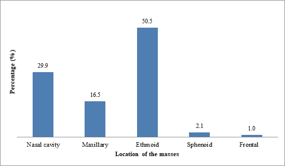
Commonest anatomical origin of sinonasal masses was ethmoid sinuses in 49 (50.5%) followed by nasal cavity in 29 (29.9%) and maxillary sinuses in 16 (16.5%) as illustrated in Figure 1.
The main clinical features were nasal blockage 81 (83.5%), nasal discharge 62 (63.9%), headache 51 (52.6%), epistaxis 41 (42.3%), bouts of sneezing 34 (35.1%) and hyposmia/anosmia 26 (26.8%) as demonstrated in Table 4.
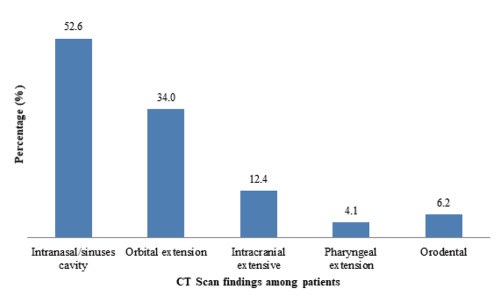
Distribution of Sinonasal masses extension into surrounding organ were were as follow: 51 (52.6%) Intranasal/paranasal sinuses, 33 (34.0%) orbital extension, 12 (12.4%) intracranial extension and 6 (6.2%) orodental extension (Figure 2).
The histological diagnosis in this study was ethmoidal (simple) nasal polyps in 48 (49.5%), squamous cell carcinoma in 17 (17.5%), antrochoanal polyp in 9 (9 3%), inverted papilloma in 9 (9.3%) and adenocarcinoma in 6 (6.2%). Histological diagnosis of the sinonasal masses showed simple inflammatory nasal polyp in 57 (58.8%), a benign tumour in 13 (13.4%) and malignant tumour in 23 (23.7%) as seen in Table 5.
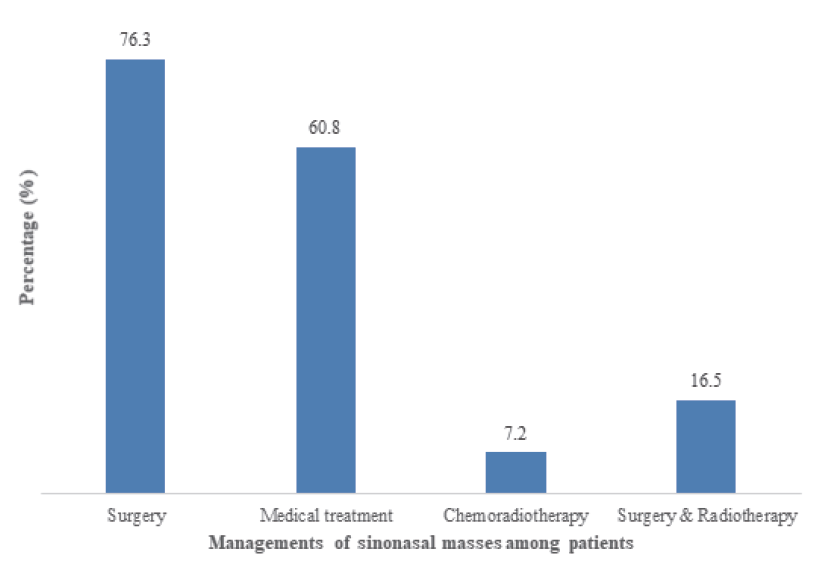
In this study, patients were managed by 74 (76.3%) surgery, 16 (16.5%) surgery and radiotherapy and 7 (7.2%) chemoradiotherapy as in figure 3. Recurrent sinonasal masses are and death from sinonasal malignancy were 8 (8.2%) and 2 (2.1%) respectively.
|
|
|
|
|
|
|
|
|
|
DISCUSSION |
|---|
Sinonasal masses arise from the nasal cavity and paranasal sinuses lined stratified squamous, respiratory-type pseudo-stratified columnar, and transitional (intermediate) epithelium (8). The prevalence in this study was high as noted in previous study (9).
In this study, the peak prevalence of sinonasal masses presented at 2nd to 4th decade of life as observed in previous report (10, 11). There was male dominance over female as reported in other studies (10). This may be due to male members are exposed to varied environmental stress and hazard factors in the course of earning a livelihood for the family and overall higher possibility of male attendance at hospitals. Female dominance was reported in other studies (12).
The effects of family and social history were observed in our study which was similar to report from other studies (13). Majority of the patients were urban dwellers while rural dwellers presented with advanced cases, this may be due to accessibility to the health facilities located in the state capital. Education, religion, marital status and occupation did not have risk factors on sinonasal masses from our study as in previous study (11).
Majority of the patients presented late after 3 months with advanced sinonasal masses which may be due to hidden nature of the masses, misdiagnosis of cases as flu until the masses are obvious or complicated as in previous report (11). Unilateral conditions occurred from tumour and antrochoanal polyps are commoner than bilateral cases secondary to ethmoidal (simple) nasal polyps as in other studies (14). Recurrent sinonasal masses are very common with nasal polyps and malignant tumour in this study which is similar to report from other studies (15). In this study, all sinonasal masses are solitary except simple nasal polyps.
The most common presenting clinical features in the present research work were nasal obstruction and nasal discharge and headache similar to report from other studies (16-18). Other findings include epistaxis mainly from neoplastic masses, large sinonasal masses causes different ranges of loss of smell and voice changes from hyponasality (19). Symptoms of upper airway allergy-like rhinorrhea, bouts of excessive sneezing were also noticed in some patients with nasal polyps and these support the fact that allergy plays a major role in nasal polyp (19). Allergic test to confirm allergic disorder was not documented in the patient’s record in this study.
Clinical examination in this study revealed sinonasal masses arises from both the nasal cavity and paranasal sinuses. Nasal Polyps are commoner than the neoplastic tumour. The commonest origin was ethmoid sinuses this may be because polyps are the most common causes of sinonasal masses in our findings. Further examination revealed the majority of the unilateral sinonasal masses were neoplastic. Microscopic examination of unilateral sinonasal masses is required to rule out neoplastic changes with malignancy in particular as it was depicted from other studies (19-21). Diagnostic nasal endoscopy is an advanced diagnostic tool did not help in these patients due to late presentation and distorted anatomy contrary to findings in other studies (18).
Computerized tomography (CT) scan of the paranasal sinuses and base of the skull was performed on selected patients to determine the type, extent and changes in the sinonasal tract by benign masses also in malignancy to also detect the expansion, bone remodelling, aggressive destruction and invasion of adjacent tissues, causing ill-defined margins and organs (7). In this study, it reveals the extent of the masses to be nasal and paranasal sinuses as the commonest invasion. Due to late presentation of the patients, most of the sinonasal masses have extended into the surrounding organ such as orbit, cranium and dentopalatine region. This is to prepare and avoid dangerous complications from orbit and cranium.
Intraoperative and histological examination revealed that the observed sinonasal masses to be polyps, neoplastic tumour and infective pathology. The infected masses were fungal mass and frontoethmoidal mucocele. Non-neoplastic masses were commoner than neoplastic masses and the commonest non neoplastic masses was polyps which is similar to what was reported by other studies (22, 23). Further analysis of the neoplastic sinonasal masses of our study revealed malignancy to be commoner than benign masses this is contrary to findings in other studies (24). The commonest benign sinonasal masses was inverted papilloma in this study while Haemangiomas was the commonest in some reported studies (18, 24). The most common malignant sinonasal mass in our findings was squamous cell carcinoma which is similar to records from Nepal (25).
Treatment offered to these patients were surgeries (Intranasal polypectomy, Partial or total maxillectomy, etc) medical treatment, radiotherapy, chemoradiotherapy, and outpatient followed up in different combination. Sinonasal polyps had surgical excision done. Polyps were further treated with antihistamine and steroid nasal spray while fungal sinonasal masses were treated with systemic antifungal drugs. Benign sinonasal masses were offered excision with a wide margin. Early stages malignancy had wide surgical excision. Malignant sinonasal masses in advanced stage had an excisional biopsy and referred for chemoradiation. All our patients had regular follow-up medical review in the ear, nose and throat department outpatient clinic to detect early recurrence (26). One case of recurrent malignant sinonasal masses were recorded in this study. Two deaths were recorded from advanced sinonasal malignancy during chemoradiotherapy.
|
|
LIMITATIONS |
|---|
1. It is hospitable based research and may not truly represent the whole community/country;
2. It was not all patients with the sinonasal tumors that consented for the study.
|
|
CONCLUSION |
|---|
Sinonasal masses are perceived and presented as a simple disorder with nasal obstructions and discharge. It consists of polyps and malignant tumour. Nasal Polyps are commoner than the neoplastic tumour. The commonest origin of the sinonasal masses was ethmoid sinuses which may be because polyps are the most common causes of sinonasal masses as shown in our study. Further evaluation revealed that majority of the unilateral sinonasal masses were neoplastic. They are poorly managed and presented in advanced stage to the otorhinolaryngologist, head and neck surgeon. Late presented patients had palliative treatment with resultant high recurrence and fatality. Thus Health education, serial and early screening are highly recommended.
|
|
FUNDING |
|---|
There was no financial support. It is a self-sponsored research study.
|
|
CONFLICT OF INTERESTS |
|---|
All the authors declare that there was no competing interest.
|
|
ETHICAL CONSIDERATION |
|---|
Ethical approval was obtained from the ethics and research committee of our institution.
|
|
ACKNOWLEDGMENTS |
|---|
The authors are most grateful to Ekiti state university teaching hospital, the staff and all the patients who participated in this study.
|
|
REFERENCES |
|---|