Received- August 21, 2018; Accepted- November 1, 2018
International Journal of Biomedical Science
14(2), 85-88, Dec 15, 2018
© 2018 Sanjay M. Khaladkar et al. Master Publishing Group
Neurovascular Lipomatous Hamartoma in scapular region
Sanjay M. Khaladkar, Aarushi Gupta, Shibani Saluja, Ronak Savani, Radhika Jaipuria
Department of Radiodiagnosis, Dr. D.Y.Patil Medical College and Research Center, Dr.D.Y.Patil Vidyapeeth, Pimpri, Pune India
Corresponding Author: Dr. Sanjay M. Khaladkar, Professor, Department of Radiodiagnosis, Dr. D.Y.Patil Medical College and Research Center, Dr.D.Y.Patil Vidyapeeth, Pimpri, Pune India. E-mail: drsanjaymkhaladkar@gmail.com.
|
|
ABSTRACT
 |
Hamartoma is defined as non-neoplastic malformation characterized by proliferation of mature cells and tissues indigenous to the affected part. Excess of one or more tissues in a disorganized manner can occur. Usually hamartomas are present at birth or in young age but also reported in later ages of life. Hence diagnosis of hematomas cannot be made on the age factor. No diagnostic criteria have been laid down for Neurovascular Hamartoma (NVH). NVH contain small to medium sized vessels and closely packed groups of well-formed nerve bundles in loose connective tissue matrix. NMH are rare intra-neural hamartomas having admixture of mature neural elements, mature skeletal elements and no cellular atypia. We report a case of 25-year-old male patient with swelling in left scapular region since 12 years. On imaging, it was diagnosed as neurovascular lipomatous hamartoma due to admixture of neural elements, fat and vessels.
KEY WORDS:
Hamartoma; nerve hamartoma; neuromuscular hamartoma; neurovascular lipomatous hamartoma
|
|
INTRODUCTION |
The term hamartoma was first introduced by Albrecht in 1904. It was derived from a Greek word “Hamartia” which means a character flaw (that is a failure). He described Hamartoma as an error of tissue development or tumour like non-neoplastic malformations which results from abnormal admixture of tissue indigenous to the site (1). Hamartoma, choristoma, teratoma and dermoid are different histologically. A hamartoma is composed of abnormal proliferation of tissue endogenous to involved site. Choristoma consists of abnormal proliferation of tissues exogenous to occurring site. Teratoma consists of pluripotent tissues which are foreign to site of occurrence. Pluripotent tissue is derived from all the germ layers. Dermoids are usually cystic and contain either ectodermal and mesodermal elements, usually the former (2).
Teratomas have the highest malignant transformation rate. Hamartomas have little or no malignant transformation rate while choristomas have less than 1% chance of malignant transformation. Hamartomas commonly occur in skin, chest wall, lungs, liver, gastrointestinal tract and kidneys. According to the predominance of tissue, they can be classified as lipomatous, neurogenic, vascular, angiomatous, osseous and chondroid (2).
|
|
CASE REPORT |
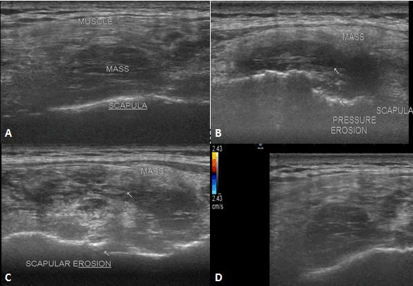
A 25- year old male patient presented with swelling in the left scapular region since 12 years. Initially, it was small in size, which gradually increased in size. Local Ultrasonography (USG) done with a linear probe (7-12 MHz) showed a large irregularly marginated heterogeneously hypoehoic solid mass of size approximately 11 × 8 × 4 cms in transverse, cranio-caudal and antero-posterior dimensions respectively, in relation to the posterior surface of body of scapula and its infraspinous portion showing no significant vascularity on colour Doppler. It was causing pressure erosions on the underlying body of scapula (Figure 1).
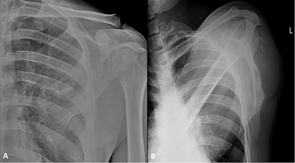
A radiograph of chest AP view and left scapula AP view showed osteolytic lesions in infraspinous portion of body of scapula with subtle sclerotic margins (Figure 2).
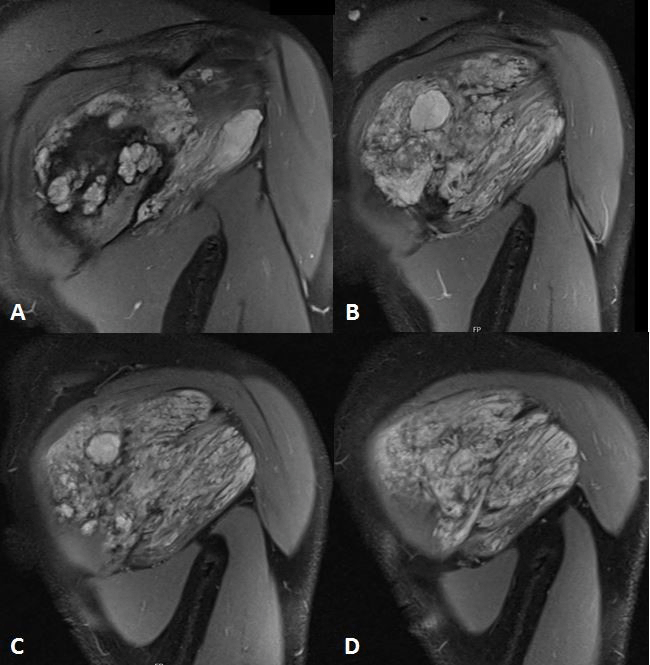
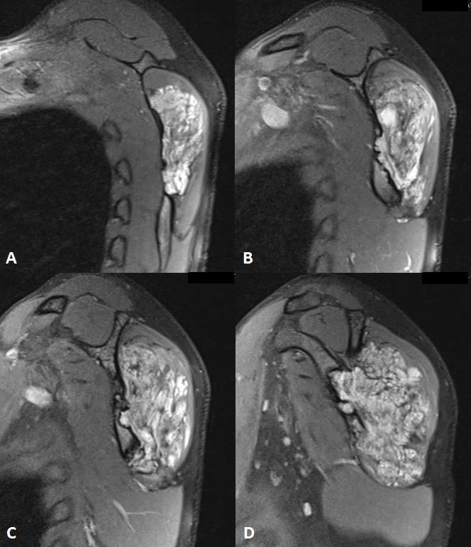
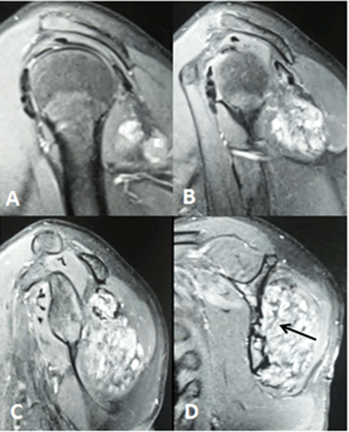
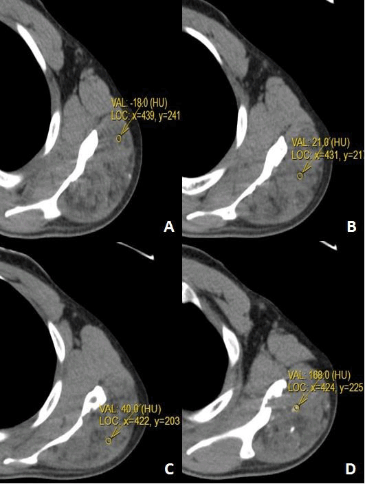
MRI of left shoulder showed a large well defined mass with lobulated outlines measuring approximately 11.6 × 8.2 × 4.3 cms in transverse, cranio-caudal and antero-posterior dimensions respectively in relation to posterior surface of the infraspinous portion of body of the scapula. It was causing posterior displacement and involvement of adjacent infraspinatus muscle. It was slightly hyperintense with respect to muscle on T1WI (Figure 3) and heterogeneously hyperintense on T2WI (Figure 3) and PDFS (Figure 4 and 5). It showed multiple septations appearing hypointense on T2WI. A cystic area measuring approx. 2.3 × 1.8 cms was noted in its medial portion appearing hypointense on T1WI and hyperintense on T2WI and PDFS with fluid- fluid levels – suggestive of cystic degeneration. It showed multiple foci of fat appearing hyperintense on T1WI and T2WI, nullified on PDFS – suggestive of fat components. GRE T2WI (Figure 6) showed foci of blooming suggestive of calcification. Contrast study showed avid enhancement. A diagnosis of neurovascular lipomatous hamartoma was made, most probably arising from suprascapular nerve.
CT showed multiple foci of fat and multiple nodular areas of calcification within the soft tissue mass causing pressure erosion on underlying scapula. (Figure 7 and 8)
A fragmented biopsy from peri-scapular mass comprised of soft tissue, mature adipose tissue, fibrin, pseudocystic pattern with prominent blood vessels and nerve. On immunohistochemistry, S 100 protein highlighted interspersed neural tissue. AE1/AE3 was negative. CD-34 highlighted interspersed blood vessels. Biopsy was suggestive of a benign vascular lesion/ hamartoma. An excision biopsy was suggested which was refused by the patient.
View larger version :
[in a new window] |
Figure 1. Local ultrasound of left scapular region with linear 8-12 MHz probe showed a large solid mixed echoic mass along dorsal aspect of infraspinous portion of body of scapula causing pressure erosions on underlying scapula showing no significant vascularity on colour Doppler (A-D).
|
|
View larger version :
[in a new window] |
Figure 2. Radiograph of left scapula AP and tangential view shows soft tissue swelling with multiple calcific foci / Phleboliths on dorsal aspect of scapular body with multiple osteolytic lesions due to pressure erosions.
|
|

View larger version :
[in a new window] |
Figure 3. MRI left scapula (coronal T1WI – A, B and coronal T2WI – C, D) shows a large well defined mass with lobulated outlines in relation to posterior surface of infraspinous portion of body of scapula showing fat, soft tissue and cystic components.
|
|
View larger version :
[in a new window] |
Figure 4. MRI left scapula (coronal PDFS) shows a large well defined mass with lobulated outlines in relation to posterior surface of infraspinous portion of body of scapula showing fat, soft tissue and cystic components causing marked pressure erosion on adjoining scapular body.
|
|
View larger version :
[in a new window] |
Figure 5. MRI left scapula (sagittal PDFS) shows a large well defined mass with lobulated outlines in relation to posterior surface of infraspinous portion of body of scapula showing fat, soft tissue and cystic components causing marked pressure erosion on adjoining scapular body.
|
|

View larger version :
[in a new window] |
Figure 6. MRI left scapula (axial GRE T2) shows a large well defined mass with lobulated outlines in relation to posterior surface of infraspinous portion of body of scapula showing blooming within the mass due to calcification causing marked pressure erosion on adjoining scapular body.
|
|
View larger version :
[in a new window] |
Figure 7. Plain CT scan of thorax shows a well defined mass on dorsal aspect of infraspinous portion of body of scapula showing soft tissue, fat components and multiple nodular calcific foci/ Phleboliths.
|
|
View larger version :
[in a new window] |
Figure 8. Post contrast sagittal T1FS (T1 weighted fat saturated) (A-D) showing intense enhancement (marked by black arrow in C and D) in vascular component of neurovascular hamartoma.
|
|
|
|
DISCUSSION |
Hamartoma is defined as non-neoplastic malformation characterized by proliferation of mature cells and tissues indigenous to the affected part (3). Excess of one or more tissues in a disorganized manner can occur. Usually hamartomas are present at birth or in young age but also reported in later ages of life. Hence diagnosis of hematomas cannot be made on the age factor. No diagnostic criteria have been laid down for Neurovascular Hamartoma (NVH).
Histopathologically, differential diagnosis for NVH is haemangioma. Haemangiomas usually are present at birth and rapidly proliferate in infancy and involute in early childhood. They lack or show minimal neural component.
Hamartomas are usually asymptomatic, even if present at birth and may not be apparent until adolescence or adulthood. They show both neural and vascular components. Histopathologically, NVH contain small to medium sized vessels and closely packed groups of well-formed nerve bundles in the loose connective tissue matrix. There is no inflammatory component (2).
Nerve hamartomas are rare and often clinically silent. They are usually misdiagnosed.
Nerve hamartomas in peripheral nervous systems are either fibrolipomatous (FLH) or neuromuscular hematomas (NMH). FLH (intraneural lipoma, perineural lipoma, lipomatous hamartoma) are fibrofatty malformations of peripheral nerve which are rare. They occur due to overgrowth of fibrous adipose tissue within and surrounding nerve bundles causing nerve enlargement. They occur in the first three decade of life and can occur in digital, ulnar, radial, sciatic, peroneal, cranial nerves and brachial plexus. Histologically, they are composed of mature adipose and connective tissue admixed with the nerve fibres. NMH are rare intra-neural hamartomas having an admixture of mature neural elements, mature skeletal elements and no cellular atypia. They arise from one of the four mechanisms - skeletal muscle entrapment within in the nerve during embryogenesis, muscle spindle hamartomas growth, neuro-ectodermal differentiation into skeletal muscle or epigenetic alteration of motor end plates (4).
|
|
CONCLUSION |
Neurovascularlipomatous hamartomas are rare benign tumours arising from the nerve. High index of suspicion is needed for its diagnosis on imaging. Presence of fat, soft tissue, vascular component, phleboliths in varying amounts is diagnostic. Pressure erosion of adjoining bone can occur due to long term compression.
|
|
CONFLICT OF INTEREST |
The authors declare that no conflicting interests exist.
|
|
REFERENCES |
- Albretht E. Üeber hamartome. Verh Dtsch Ges Pathol. 1904; 7: 153-157.
- Fande PZ, Patil SK, Gadbail AR, Ghatage DD, et al. Neurovascular Hamartoma of Face: An Unusual Clinical Presentation. World J. Dent. 2017; 8 (2): 151-154.
- Mannan AA, Sharma MC, Singh MK, Bahadur S, et al. Vascular hamartoma of the paranasal sinuses: report of 3 rare cases and a short review of the literature. Ear Nose Throat J. 2009 Jan; 88 (1): 740-743.
- Senger JL, Kanthan R. Nerve Hamartomas - Fact or Fiction? Surg Oncol Clin Pract J. 2016; 1 (1): 1004.